

ImmunoPET of tissue factor expression in triple-negative breast cancer with a radiolabeled antibody Fab fragment
- PMID: 25801992
- PMCID: PMC4482783
- DOI: 10.1007/s00259-015-3038-1
ImmunoPET of tissue factor expression in triple-negative breast cancer with a radiolabeled antibody Fab fragment
Abstract
Purpose: To date, there is no effective therapy for triple-negative breast cancer (TNBC), which has a dismal clinical outcome. Upregulation of tissue factor (TF) expression leads to increased patient morbidity and mortality in many solid tumor types, including TNBC. Our goal was to employ the Fab fragment of ALT-836, a chimeric anti-human TF mAb, for PET imaging of TNBC, which can be used to guide future TNBC therapy.
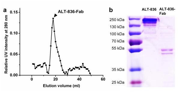
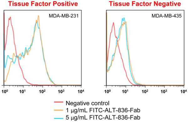
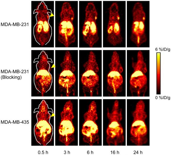
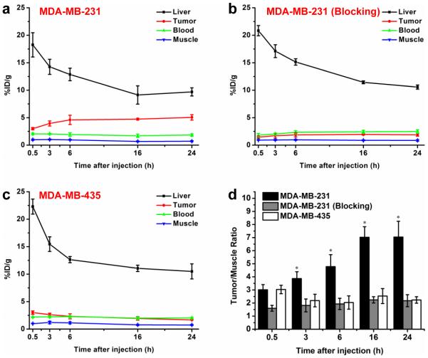
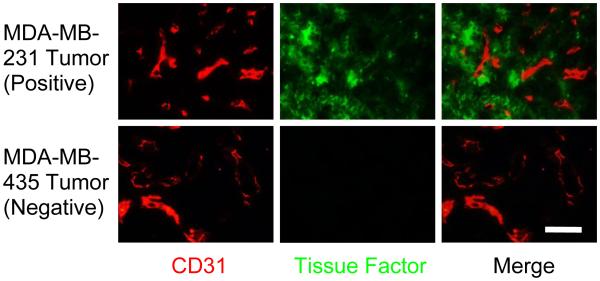
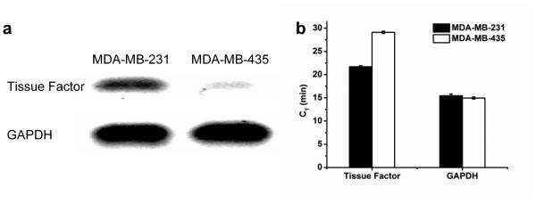
Methods: ALT-836-Fab was generated by enzymatic papain digestion. SDS-PAGE and FACS studies were performed to evaluate the integrity and TF binding affinity of ALT-836-Fab before NOTA conjugation and (64)Cu-labeling. Serial PET imaging and biodistribution studies were carried out to evaluate the tumor targeting efficacy and pharmacokinetics in the MDA-MB-231 TNBC model, which expresses high levels of TF on the tumor cells. Blocking studies, histological assessment, as well as RT-PCR were performed to confirm TF specificity of (64)Cu-NOTA-ALT-836-Fab.
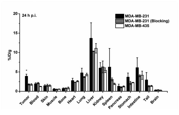
Results: ALT-836-Fab was produced with high purity, which exhibited superb TF binding affinity and specificity. Serial PET imaging revealed rapid and persistent tumor uptake of (64)Cu-NOTA-ALT-836-Fab (5.1 ± 0.5 %ID/g at 24 h post-injection; n = 4) and high tumor/muscle ratio (7.0 ± 1.2 at 24 h post-injection; n = 4), several-fold higher than that of the blocking group and tumor models that do not express significant level of TF, which was confirmed by biodistribution studies. TF specificity of the tracer was also validated by histology and RT-PCR.
Conclusion: (64)Cu-NOTA-ALT-836-Fab exhibited prominent tissue factor targeting efficiency in MDA-MB-231 TNBC model. The use of a Fab fragment led to fast tumor uptake and good tissue/muscle ratio, which may be translated into same-day immunoPET imaging in the clinical setting to improve TNBC patient management.
Figures
Similar articles
-
PET imaging of CD105/endoglin expression with a ⁶¹/⁶⁴Cu-labeled Fab antibody fragment.Eur J Nucl Med Mol Imaging. 2013 May;40(5):759-67. doi: 10.1007/s00259-012-2334-2. Epub 2013 Jan 24. Eur J Nucl Med Mol Imaging. 2013. PMID: 23344138 Free PMC article.
-
ImmunoPET imaging of Trop2 expression in triple-negative breast cancer using [64Cu]Cu-NOTA-Trodelvy-F(ab')2.Eur J Nucl Med Mol Imaging. 2025 Jul;52(9):3223-3237. doi: 10.1007/s00259-025-07167-5. Epub 2025 Feb 25. Eur J Nucl Med Mol Imaging. 2025. PMID: 39994021
-
ImmunoPET and Near-Infrared Fluorescence Imaging of Pancreatic Cancer with a Dual-Labeled Bispecific Antibody Fragment.Mol Pharm. 2017 May 1;14(5):1646-1655. doi: 10.1021/acs.molpharmaceut.6b01123. Epub 2017 Mar 24. Mol Pharm. 2017. PMID: 28292180 Free PMC article.
-
64Cu-1,4,7-Triazacyclononane-1,4,7-triacetic acid-p-isothiocyanatobenzyl-ALT-836.2012 Dec 6 [updated 2013 Mar 21]. In: Molecular Imaging and Contrast Agent Database (MICAD) [Internet]. Bethesda (MD): National Center for Biotechnology Information (US); 2004–2013. 2012 Dec 6 [updated 2013 Mar 21]. In: Molecular Imaging and Contrast Agent Database (MICAD) [Internet]. Bethesda (MD): National Center for Biotechnology Information (US); 2004–2013. PMID: 23534079 Free Books & Documents. Review.
-
Copper-64-immunoPET imaging: bench to bedside.Q J Nucl Med Mol Imaging. 2020 Dec;64(4):356-363. doi: 10.23736/S1824-4785.20.03310-5. Epub 2020 Oct 12. Q J Nucl Med Mol Imaging. 2020. PMID: 33045821 Review.
Cited by
-
Recent advances in nanotheranostics for triple negative breast cancer treatment.J Exp Clin Cancer Res. 2019 Oct 28;38(1):430. doi: 10.1186/s13046-019-1443-1. J Exp Clin Cancer Res. 2019. PMID: 31661003 Free PMC article. Review.
-
PET imaging of Aspergillus infection using Zirconium-89 labeled anti-β-glucan antibody fragments.Eur J Nucl Med Mol Imaging. 2024 Sep;51(11):3223-3234. doi: 10.1007/s00259-024-06760-4. Epub 2024 May 24. Eur J Nucl Med Mol Imaging. 2024. PMID: 38787397 Free PMC article.
-
Advances in immunoPET/SPECT imaging: The role of Fab and F(ab')2 fragments in theranostics.Acta Pharm Sin B. 2025 Aug;15(8):3888-3924. doi: 10.1016/j.apsb.2025.05.030. Epub 2025 May 29. Acta Pharm Sin B. 2025. PMID: 40893670 Free PMC article. Review.
-
Copper-64 Labeled PEGylated Exosomes for In Vivo Positron Emission Tomography and Enhanced Tumor Retention.Bioconjug Chem. 2019 Oct 16;30(10):2675-2683. doi: 10.1021/acs.bioconjchem.9b00587. Epub 2019 Sep 27. Bioconjug Chem. 2019. PMID: 31560538 Free PMC article.
-
Radioimmunotherapy with an 211 At-labeled anti-tissue factor antibody protected by sodium ascorbate.Cancer Sci. 2021 May;112(5):1975-1986. doi: 10.1111/cas.14857. Epub 2021 Mar 30. Cancer Sci. 2021. PMID: 33606344 Free PMC article.
References
-
- Foulkes WD, Smith IE, Reis-Filho JS. Triple-negative breast cancer. New Engl J Med. 2010;363:1938–48. - PubMed
-
- Terwisscha van Scheltinga AG, Berghuis P, Nienhuis HH, Timmer-Bosscha H, Pot L, Gaykema SB, et al. Visualising dual downregulation of insulin-like growth factor receptor-1 and vascular endothelial growth factor-A by heat shock protein 90 inhibition effect in triple negative breast cancer. Eur J Cancer. 2014;50:2508–16. - PubMed
-
- Carey L, Winer E, Viale G, Cameron D, Gianni L. Triple-negative breast cancer: disease entity or title of convenience? Nat Rev Clin Oncol. 2010;7:683–92. - PubMed
-
- Callander NS, Varki N, Rao LV. Immunohistochemical identification of tissue factor in solid tumors. Cancer. 1992;70:1194–201. - PubMed
-
- Vrana JA, Stang MT, Grande JP, Getz MJ. Expression of tissue factor in tumor stroma correlates with progression to invasive human breast cancer: paracrine regulation by carcinoma cell-derived members of the transforming growth factor beta family. Cancer Res. 1996;56:5063–70. - PubMed
Publication types
MeSH terms
Substances
Grants and funding
LinkOut - more resources
Full Text Sources
Other Literature Sources
Miscellaneous
