. 2015 Apr 27;33(18):2167-74.
doi: 10.1016/j.vaccine.2015.02.067. Epub 2015 Mar 21.

Recombinant rubella vectors elicit SIV Gag-specific T cell responses with cytotoxic potential in rhesus macaques

Affiliations

Recombinant rubella vectors elicit SIV Gag-specific T cell responses with cytotoxic potential in rhesus macaques

Margherita Rosati et al. Vaccine. .

Abstract

Live-attenuated rubella vaccine strain RA27/3 has been demonstrated to be safe and immunogenic in millions of children. The vaccine strain was used to insert SIV gag sequences and the resulting rubella vectors were tested in rhesus macaques alone and together with SIV gag DNA in different vaccine prime-boost combinations. We previously reported that such rubella vectors induce robust and durable SIV-specific humoral immune responses in macaques. Here, we report that recombinant rubella vectors elicit robust de novo SIV-specific cellular immune responses detectable for >10 months even after a single vaccination. The antigen-specific responses induced by the rubella vector include central and effector memory CD4(+) and CD8(+) T cells with cytotoxic potential. Rubella vectors can be administered repeatedly even after vaccination with the rubella vaccine strain RA27/3. Vaccine regimens including rubella vector and SIV gag DNA in different prime-boost combinations resulted in robust long-lasting cellular responses with significant increase of cellular responses upon boost. Rubella vectors provide a potent platform for inducing HIV-specific immunity that can be combined with DNA in a prime-boost regimen to elicit durable cellular immunity.

Keywords: CM9 tetramer response; Cytotoxic T cells; DNA prime rubella boost; DNA vaccine; De novo cellular responses; HIV vaccine; IFN-γ response; Live attenuated viral vector; Memory T cells; Recall cellular responses; Rhesus macaque; Rubella prime boost; Rubella vaccine strain RA27/3; SIV Gag.


Conflict of interest statement

Conflict of interest: GNP and BKF are inventors on US Government-owned patents and patent applications related to DNA vaccines and gene expression optimization. N.Y.S. is a full time employee of Inovio Pharmaceuticals and as such receives compensation in the form of salary and stock options. There are no further patents, products in development or marketed products to declare. This does not alter our adherence to all the policies on sharing data and materials. The remaining authors declare that no other competing interests exist.

Figures

FIGURE 1.
FIGURE 1.. Vaccination protocols.
The cartoon is an adaptation from Virnik et al. [19] and details the vaccination timeline used for this Indian rhesus macaque study. Animals CL6A and CL6J had received RA27/3 rubella vaccine strains and sham DNA. Macaques CL67, CL49, CL6V had received rubella type 1 and type 2 which were “no takes” and did not induce antibodies to rubella and also did not induce Gag-specific T cell immunity. The animals received sequential vaccinations with DNA and rubella vector type 3 or type 3*, referred to as rubella vector herein, expressing SIV Gag. The animals were analyzed for Gag-specific IFN-γ+ T cell responses, and macaques positive for the MamuA*01 haplotype were also analyzed for Gag CM9 tetramer responses.
FIGURE 2.
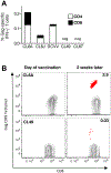
FIGURE 2.. Recombinant rubella vector vaccination induces de novo Gag-specific responses in macaques.
(A) CD4+ and CD8+ Gag-specific IFN-γ+ T cell responses measured 2 weeks after vaccination with the rubella vector in five animals (CL6A, CL6J, DCVV, CL49, CL67). (B) Dot plots show CM9 Gag tetramer responses in MamuA*01+ macaques (CL6A and CL49) at the day of vaccination and 2 weeks later.
FIGURE 3.
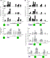
FIGURE 3.. Responses after different prime boost regimens with rubella vector and gag DNA.
(A) DNA vaccination of rubella vector primed macaques, and a 2nd rubella vector boost. The Gag-specific IFN-γ+ T cell responses (upper panels) of macaques CL67, CL49, and DCVV and the Gag CM9 tetramer-specific CD8+ T cells of the MamuA*01+ CL49 (lower panel) were measured at the indicated time points. The data from weeks 18 and 20 are from Figure 2A. (B) Rubella vector boosts of DNA-primed macaques. The Gag-specific IFN-γ+ T cells of J6L, V584 and V200 (top panels), and the CM9 tetramer-specific CD8+ T cells of the MamuA*01+ J6L, V584 and CL6V (lower panels) were measured at the indicated time points. CL6V could not be analyzed for Gag-specific IFN-γ+ T cell responses due to high background levels in the unstimulated PBMC at all time points. (C) Gag-specific responses of two macaques (CL6A, CL6J) after vaccination with rubella vector only. The Gag-specific IFN-γ+ T cell responses (black bars) are shown for both macaques, and the CM9 tetramer responses (grey bars) are shown for the MamuA*01+ CL6A. Data after the 1st rubella vector vaccination are from Figure 2. (D) Changes in cellular immunity upon a 2nd rubella vector vaccination. Gag-specific IFN-γ+ T cells (left panel) and CM9 tetramer-specific CD8+ T cells (right panel) are shown at day of vaccination and 2 weeks later. P values were obtained using paired t test. Left panel includes data from 8 macaques shown in Figures 3A (CL49, CL67, DCVV; weeks 100–102), 3B (J6L, V584, V200, CL6V (weeks 57–59) and 3C (CL6A, CL6J; weeks 100–102). Right panel includes data from 5 macaques depicted in Figures 3A (CL49; weeks 100–102), 3B (J6L, V584, CL6V (weeks 57–59) and 3C (CL6A; weeks 100–102). (E) Changes in cellular immunity in DNA-primed macaques upon rubella vector boost comparing Gag-specific IFN-γ+ T cells (left panel) and CM9 tetramer-specific CD8+ T cells (right panel) responses at day of vaccination and 2 weeks after rubella vector boost. P values were obtained using paired t test. Left panel includes data from 6 macaques shown in Figures 3A (CL49, CL67, DCVV; weeks 100–102) and 3B (J6L, V584, V200 (weeks 27). Right panel includes data from 4 macaques shown in Figures 3A (CL49; weeks 100–102) and 3B (J6L, V584, at weeks 25–27, and CL6V at weeks 100–102).
FIGURE 4.
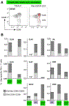
FIGURE 4.. Phenotypic analysis of Gag-specific memory T cells.
Gag-specific responses were analyzed by flow cytometry using an antibody cocktail for memory markers. (A) Dot plot overlays show the phenotypic characterization (CM-like: CD95+CD28+; effector memory EM: CD95+CD28) of the Gag-specific T cells induced by rubella vector type 3 vaccination in macaque DCVV at the day of vaccination and 2 weeks later. (B) The percentage of Gag-specific IFN-γ+ T cells with CM-like and EM cells are shown 2 weeks after the indicated vaccinations. Responses in macaque CL6V could not be analyzed (see Fig. 3). Neg* indicates absence of detectable Gag-specific T cell responses.
FIGURE 5.
FIGURE 5.. Analysis of the cytotoxic potential of the Gag-specific T cells.
Gag-specific T cells were analyzed by flow cytometry for production of IFN-γ and the presence of granzyme B+ (GzmB) 2 weeks after the indicated vaccinations. The responses were analyzed using a Boolean gating strategy. The percentage of IFN-γ+ GzmB+ double positive antigen-specific T cells and IFN-γ+ GzmB single positive cells are shown. Neg* indicates absence of detectable Gag-specific responses, as in Figure 4.

Similar articles

Cited by

References

    1. Hansen SG, Piatak M, Jr., Ventura AB, Hughes CM, Gilbride RM, Ford JC, et al. Immune clearance of highly pathogenic SIV infection. Nature. 2013;502:100–4. - PMC - PubMed
    1. Hansen SG, Vieville C, Whizin N, Coyne-Johnson L, Siess DC, Drummond DD, et al. Effector memory T cell responses are associated with protection of rhesus monkeys from mucosal simian immunodeficiency virus challenge. Nat Med. 2009;15:293–9. - PMC - PubMed
    1. Bolton DL, Santra S, Swett-Tapia C, Custers J, Song K, Balachandran H, et al. Priming T-cell responses with recombinant measles vaccine vector in a heterologous prime-boost setting in non-human primates. Vaccine. 2012;30:5991–8. - PMC - PubMed
    1. Martins MA, Bonaldo MC, Rudersdorf RA, Piaskowski SM, Rakasz EG, Weisgrau KL, et al. Immunogenicity of seven new recombinant yellow fever viruses 17D expressing fragments of SIVmac239 Gag, Nef, and Vif in Indian rhesus macaques. PLoS One. 2013;8:e54434. - PMC - PubMed
    1. Dai K, Liu Y, Liu M, Xu J, Huang W, Huang X, et al. Pathogenicity and immunogenicity of recombinant Tiantan Vaccinia Virus with deleted C12L and A53R genes. Vaccine. 2008;26:5062–71. - PubMed

MeSH terms