. 2015 Apr 7;112(14):4489-94.
doi: 10.1073/pnas.1419228112. Epub 2015 Mar 23.

Structural and kinetic analysis of protein-aggregate strains in vivo using binary epitope mapping

Affiliations

Structural and kinetic analysis of protein-aggregate strains in vivo using binary epitope mapping

Johan Bergh et al. Proc Natl Acad Sci U S A. .

Abstract

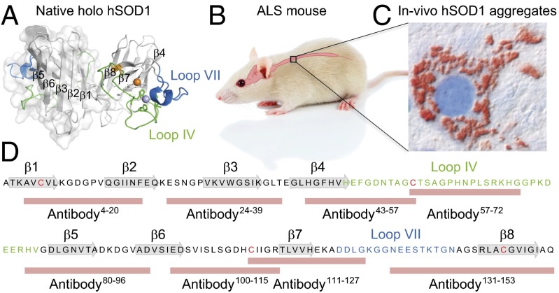
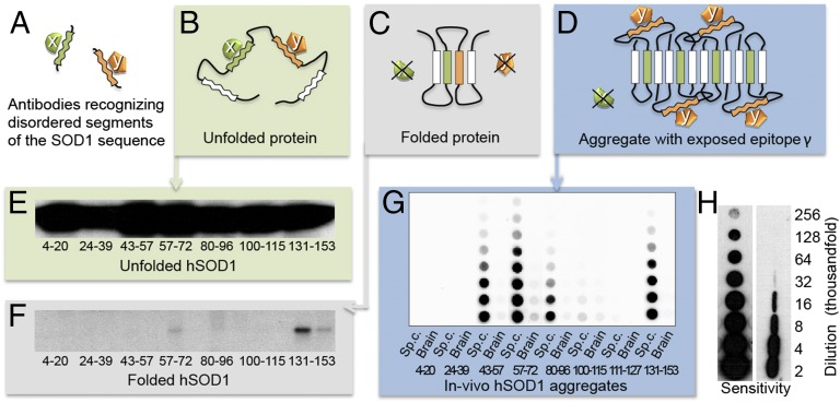
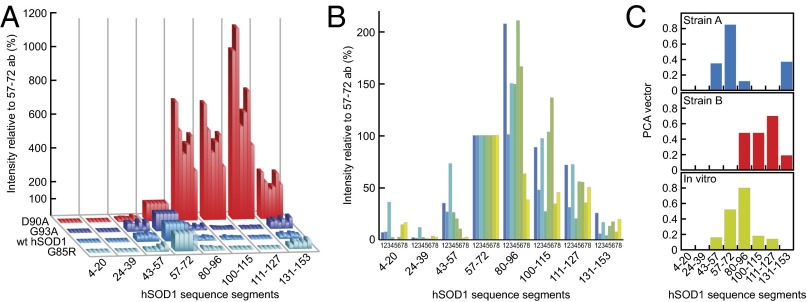
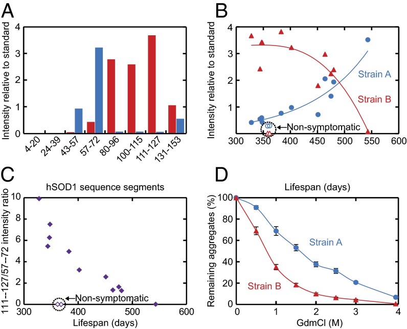
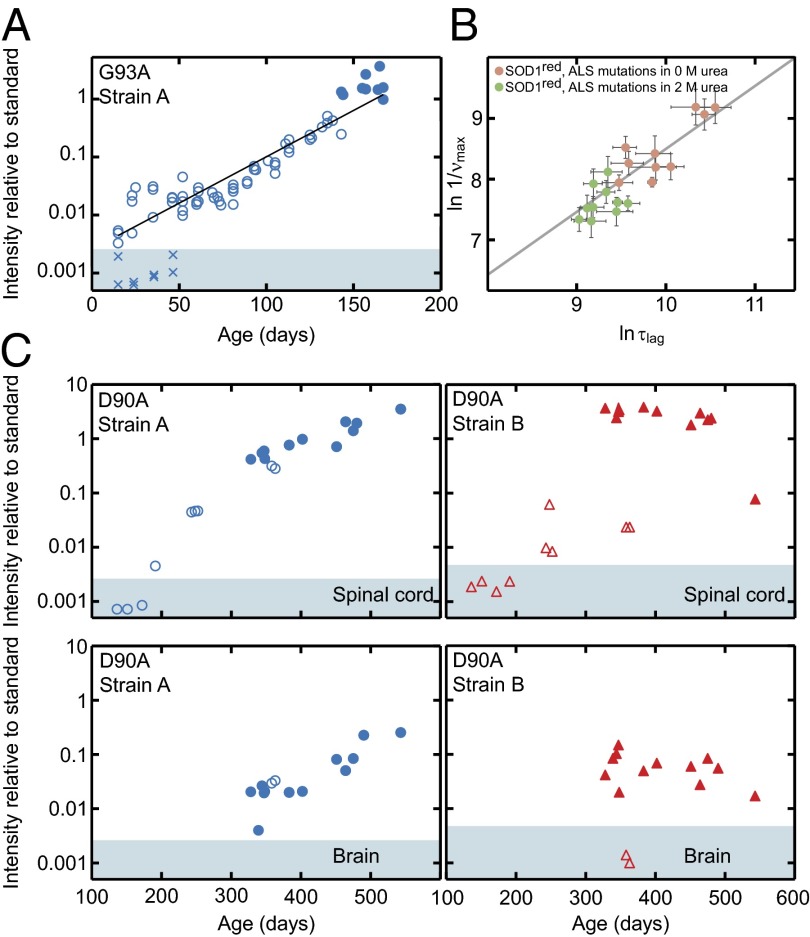
Despite considerable progress in uncovering the molecular details of protein aggregation in vitro, the cause and mechanism of protein-aggregation disease remain poorly understood. One reason is that the amount of pathological aggregates in neural tissue is exceedingly low, precluding examination by conventional approaches. We present here a method for determination of the structure and quantity of aggregates in small tissue samples, circumventing the above problem. The method is based on binary epitope mapping using anti-peptide antibodies. We assessed the usefulness and versatility of the method in mice modeling the neurodegenerative disease amyotrophic lateral sclerosis, which accumulate intracellular aggregates of superoxide dismutase-1. Two strains of aggregates were identified with different structural architectures, molecular properties, and growth kinetics. Both were different from superoxide dismutase-1 aggregates generated in vitro under a variety of conditions. The strains, which seem kinetically under fragmentation control, are associated with different disease progressions, complying with and adding detail to the growing evidence that seeding, infectivity, and strain dependence are unifying principles of neurodegenerative disease.

Keywords: amyotrophic lateral sclerosis; neurodegeneration; protein aggregation; strain; transgenic mice.


Conflict of interest statement

Conflict of interest statement: S.L.M., P.M.A., T.B., and M.O. have filed a patent partially related to concepts presented in this paper.

Figures

Fig. 1.
Fig. 1.
Human SOD1 and its involvement in the motor-neuron disease ALS. (A) Structure of the native hSOD1 homodimer showing the positions of β-strands 1–8 and loops IV and VII of the active site (PDB ID code 1HL5). (B) The pathology of transgenic mice expressing hSOD1 variants is mainly confined to the spinal cord, giving disease characteristics similar to those of human ALS. (C) Immunohistochemistry of spinal ventral horn in hSOD1G85R mouse using the 57–72 antibody showing typical accumulation of hSOD1 deposits in a motor neuron. (D) The amino acid sequence of the wild-type hSOD1 subunit, showing the positions of native secondary structure and the peptides used for generation of the antibodies used in this study.
Fig. 2.
Fig. 2.
Discrimination between “disordered” and “structured” sequence segments by the binary epitope-mapping assay. (A) Antibodies (x and y) were raised against short, consecutive segments of the hSOD1 sequence (Fig. 1). (B) In the globally unfolded state of hSOD1, the individual sequence epitopes are flexible and can adapt to the antigen-binding sites of the anti-peptide antibodies. (C) In the rigid folded hSOD1, the sequence epitopes adopt a fixed structure incompatible with antibody binding. (D) In partially ordered hSOD1 aggregates, antibody x cannot bind to its sequence epitope because this is ordered/hidden in the aggregate core, whereas antibody y can, because the epitope protrudes freely from the aggregate surface. (E) Western blot of hSOD1 captured by immobilized anti-peptide antibodies incubated with unfolded/denatured hSOD1 in solution. All of the antibodies bind, showing full exposure of flexible sequence segments in Fig. 1D. (F) Corresponding data for natively folded hSOD1, giving no binding because the sequence epitopes are now structured/hidden. Two sequential incubations were carried out, the first to capture any traces of disordered hSOD1 present in the preparation. E and F reproduced from ref. . (G) Aggregates in spinal cord and brain from a terminally ill hSOD1G93A mouse captured on a filter in a dot-blot apparatus and stained with the anti-peptide antibodies, showing the binary fingerprint of disordered and structured/hidden sequence regions of the constituent hSOD1 monomers. (H) The benchmarking 57–72 antibody detects hSOD1 in filter-captured aggregates with 10-fold higher sensitivity than hSOD1 restricted on a Western immunoblot membrane (SI Materials and Methods). The figures indicate the degree of dilution of the spinal-cord tissue.
Fig. 3.
Fig. 3.
Human SOD1 aggregation is malleable and forms two structural strains (A and B) in spinal cord, which both differ from hSOD1 fibrils produced in vitro. The data are presented normalized against the staining with the 57–72 antibody (100%) to enhance appreciation of patterns. (A) Epitope-mapping patterns of hSOD1 aggregates from terminally ill mice of the four different transgenic ALS models. Aggregate results for all hSOD1G85R, hSOD1wt, and hSOD1G93A mice presented in Table S1 are shown. The hSOD1D90A aggregates are from the six youngest mice shown in Fig. 4. (B) Epitope-mapping patterns of hSOD1 aggregates generated in vitro under eight different conditions (SI Materials and Methods). (C) Model-free PCA analysis of antibody data revealing two strains of in vivo aggregates (A and B) that differ from the hSOD1 fibrils produced in vitro (SI Materials and Methods).
Fig. 4.
Fig. 4.
Correlation between disease progression, strain levels, and their structural properties. Material from spinal-cord samples of hSOD1D90A mice. (A) Epitope-mapping patterns of hSOD1 aggregates from the youngest (328 d, red bars) and oldest (543 d, blue bars) terminally ill hSOD1D90A mice that were analyzed. (B) Coexisting strain A and B plotted against lifespans of mice. (C) Ratios of the strain-B (intensity with 111–127 antibody) and -A (intensity with 57–72 antibody) levels in B, showing clear correlation with survival time. (D) Chemical–mechanical titration of hSOD1 aggregates by sonication of spinal cord homogenates from four terminal hSOD1D90A mice in indicated concentrations of GdmCl followed by analysis in the epitope-mapping assay. Strain-B aggregates are more fragile than their strain-A counterparts.
Fig. 5.
Fig. 5.
In vivo aggregation kinetics of hSOD1 in spinal cord and brain. (A) Strain-A aggregation in spinal cord of hSOD1G93A mice shows essentially simplistic exponential growth (open and filled circles). The filled circles represent end-stage mice. X indicates brains from some of the young hSOD1G93A mice, showing absence of detectable aggregates. Brains contain as much mutant hSOD1 as spinal cords in hSOD1G93A mice (26). (B) In vitro aggregation data of monomeric reduced apo-hSOD1 variants, destabilized by single point mutations in buffer (pink circles) and 2 M urea (green circles), show a strong correlation (slope δlogτlag/δlogνmax = 1.03 ± 0.18, r = 0.88) between the apparent lag time, logτlag, and the aggregate growth rate, δlogνmax, indicating exponential kinetics analogous to the in vivo data in A. (C) Time courses of the simultaneous strain-A and -B aggregation in hSOD1D90A mice (open symbols, nonsymptomatic; closed, end-stage mice). The top panels show data from spinal cord, and the bottom panels show the matching samples from brain. The shaded areas in A and C indicate staining intensities below means + 2 SD of the results for nontransgenic controls (Table S1).

References

    1. Lu JX, et al. Molecular structure of β-amyloid fibrils in Alzheimer’s disease brain tissue. Cell. 2013;154(6):1257–1268. - PMC - PubMed
    1. Eisenberg D, Jucker M. The amyloid state of proteins in human diseases. Cell. 2012;148(6):1188–1203. - PMC - PubMed
    1. Eichner T, Radford SE. A diversity of assembly mechanisms of a generic amyloid fold. Mol Cell. 2011;43(1):8–18. - PubMed
    1. Knowles TP, et al. An analytical solution to the kinetics of breakable filament assembly. Science. 2009;326(5959):1533–1537. - PubMed
    1. Ferguson N, et al. General structural motifs of amyloid protofilaments. Proc Natl Acad Sci USA. 2006;103(44):16248–16253. - PMC - PubMed

Publication types

LinkOut - more resources