Clinical Trial
. 2015 Jun;12(6):821-30.
doi: 10.1513/AnnalsATS.201501-029OC.

Spatial Variation in the Healthy Human Lung Microbiome and the Adapted Island Model of Lung Biogeography

Affiliations
Clinical Trial

Spatial Variation in the Healthy Human Lung Microbiome and the Adapted Island Model of Lung Biogeography

Robert P Dickson et al. Ann Am Thorac Soc. 2015 Jun.

Abstract

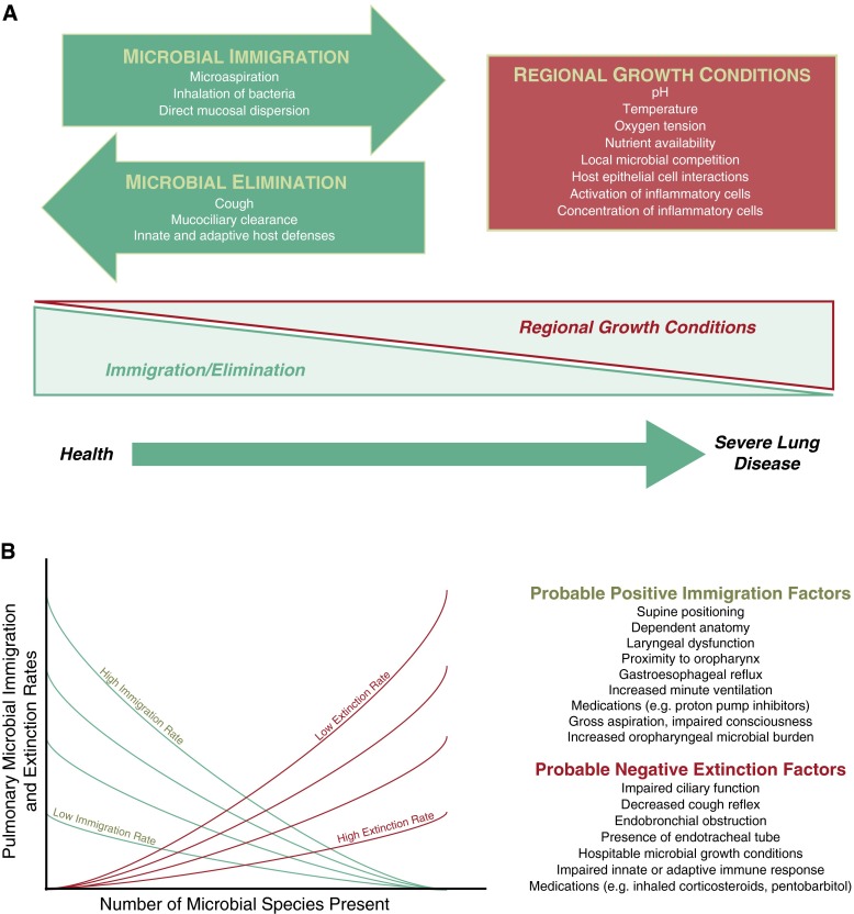
Rationale: The lung microbiome is spatially heterogeneous in advanced airway diseases, but whether it varies spatially in health is unknown. We postulated that the primary determinant of lung microbiome constitution in health is the balance of immigration and elimination of communities from the upper respiratory tract (URT; "adapted island model of lung biogeography"), rather than differences in regional bacterial growth conditions.

Objectives: To determine if the lung microbiome is spatially varied in healthy adults.

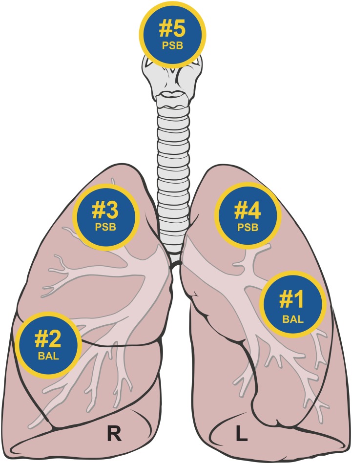
Methods: Bronchoscopy was performed on 15 healthy subjects. Specimens were sequentially collected in the lingula and right middle lobe (by bronchoalveolar lavage [BAL]), then in the right upper lobe, left upper lobe, and supraglottic space (by protected-specimen brush). Bacterial 16S ribosmal RNA-encoding genes were sequenced using MiSeq (Illumina, San Diego, CA).

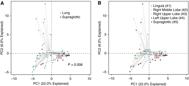
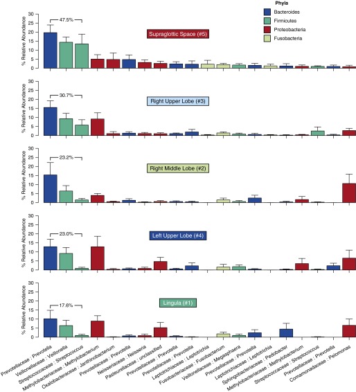
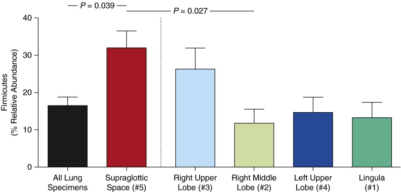
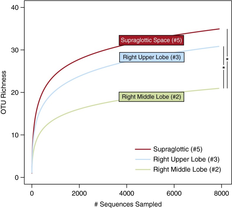
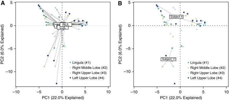
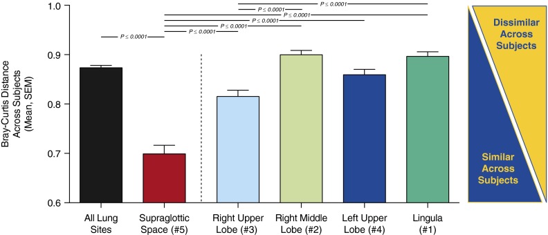
Measurements and main results: There were no significant differences between specimens collected by BAL and protected-specimen brush. Spatially separated intrapulmonary sites, when compared with each other, did not contain consistently distinct microbiota. On average, intrasubject variation was significantly less than intersubject variation (P = 0.00003). By multiple ecologic parameters (community richness, community composition, intersubject variability, and similarity to source community), right upper lobe microbiota more closely resembled those of the URT than did microbiota from more distal sites. As predicted by the adapted island model, community richness decreased with increasing distance from the source community of the URT (P < 0.05).

Conclusions: In healthy lungs, spatial variation in microbiota within an individual is significantly less than variation across individuals. The lung microbiome in health is more influenced by microbial immigration and elimination (the adapted island model) than by the effects of local growth conditions on bacterial reproduction rates, which are more determinant in advanced lung diseases. BAL of a single lung segment is an acceptable method of sampling the healthy lung microbiome. Clinical trial registered with www.clinicaltrials.gov (NCT02392182).

Keywords: 16S ribosomal DNA; bronchoscopy; lung; microbiome.


Figures

Figure 1.
Figure 1.
Ecological modeling of the respiratory microbiome. (A) The constitution of the respiratory microbiome is determined by three factors: microbial immigration, microbial elimination and the relative reproduction rates of its members. In health, community membership is primarily determined by immigration and elimination; in advanced lung disease, membership is primarily determined by regional growth conditions. Adapted with permission from Dickson and colleagues (2). (B) The adapted island model of lung biogeography. Community richness in health for a given site in the respiratory tract is a function of immigration and elimination factors. Adapted with permission from Dickson and colleagues (1).
Figure 2.
Figure 2.
Sequence of bronchoscopic sampling. After sedation and administration of local anesthetic to the upper respiratory tract, the bronchoscope was inserted through the mouth and advanced quickly and without suction to a wedge position. Bronchoalveolar lavage was performed in the lingula (#1) and then in the right middle lobe (#2). protected-specimen brushing was then performed in the right upper lobe (#3), left upper lobe (#4), and supraglottic space (#5). Figure adapted from original by Patrick J. Lynch and C. Carl Jaffe, M.D., via Creative Commons Attribution 2.5 license 2006 (http://goo.gl/xuJRCO).
Figure 3.
Figure 3.
Ordination of supraglottic and intrapulmonary bacterial communities. (A and B) Unsupervised ordination (principal components analysis) of supraglottic (SG) versus combined intrapulmonary specimens (A) or individual intrapulmonary specimens (B), labeled by specimen site. Numbers in B refer to the order of sampling during bronchoscopy. Supraglottic communities were significantly distinct from lungs collectively (P = 0.006) and, with the exception of the right upper lobe (RUL), individually (see Table 2 for significance). LUL = left upper lobe; RML = right middle lobe.
Figure 4.
Figure 4.
Relative abundance of upper respiratory tract microbiota in lung specimens. The 20 most abundant operational taxonomic units (OTUs) in supraglottic specimens are ranked in descending order of mean relative abundance and shown across specimen sites. Specimen sites are labeled by anatomic site and sequence of sampling during bronchoscopy. OTUs are labeled by family:genus. Bar plots are colored by phylum (see Phyla legend); error bars represent SEM. The sum of the three most abundant upper respiratory tract OTUs (Prevotella sp., Veillonella sp., Streptococcus sp.) is shown.
Figure 5.
Figure 5.
Relative abundance of Firmicutes phylum in spatially separated sites in the respiratory tract. Supraglottic specimens contained a significantly greater relative abundance of Firmicutes-classified operational taxonomic units than did lung specimens collectively (P = 0.04) or than right middle lobe specimens (P = 0.03; data are mean ± SEM).
Figure 6.
Figure 6.
Operational taxonomic unit (OTU) richness decreases with increased distance from the source community. Rarefaction analysis demonstrated that OTU richness was greater in the supraglottic space than in intrapulmonary sites. Richness was greater in the right upper lobe (RUL) than in the right middle lobe (RML), as predicted by the adapted island model of lung biogeography. OTU richness of the lingula (#1) and left upper lobe (#4) were intermediate between RUL and RML (see main text). Numbers refer to order of bronchoscopic sampling. *P ≤ 0.05.
Figure 7.
Figure 7.
Ordination of intrapulmonary bacterial communities. (A) Unsupervised ordination (principal components analysis) of intrapulmonary specimens, labeled by anatomic site. Numbers in key refer to order of sampling during bronchoscopy. Community membership was not significantly distinct across sites (P > 0.05 for all comparisons). (B) Principal components analysis labeled by representative subjects. Subject 11 exhibited similarity across intrapulmonary sites, whereas subject 4 exhibited heterogeneity in community membership. LUL = left upper lobe; RML = right middle lobe; RUL = right upper lobe.
Figure 8.
Figure 8.
Comparison of intersubject similarity in microbiota by anatomic site. Microbiota detected in the supraglottic space were more similar across subjects than were microbiota detected at intrapulmonary sites (P ≤ 0.001 for all). Microbiota detected in the right upper lobe were more similar across subjects than were microbiota detected in the right middle lobe or lingula (P ≤ 0.001).

References

    1. Dickson RP, Erb-Downward JR, Huffnagle GB. Towards an ecology of the lung: new conceptual models of pulmonary microbiology and pneumonia pathogenesis. Lancet Respir Med. 2014;2:238–246. - PMC - PubMed
    1. Dickson RP, Martinez FJ, Huffnagle GB. The role of the microbiome in exacerbations of chronic lung diseases. Lancet. 2014;384:691–702. - PMC - PubMed
    1. Charlson ES, Bittinger K, Haas AR, Fitzgerald AS, Frank I, Yadav A, Bushman FD, Collman RG. Topographical continuity of bacterial populations in the healthy human respiratory tract. Am J Respir Crit Care Med. 2011;184:957–963. - PMC - PubMed
    1. Morris A, Beck JM, Schloss PD, Campbell TB, Crothers K, Curtis JL, Flores SC, Fontenot AP, Ghedin E, Huang L, et al. Comparison of the respiratory microbiome in healthy nonsmokers and smokers. Am J Respir Crit Care Med. 2013;187:1067–1075. - PMC - PubMed
    1. Segal LN, Alekseyenko AV, Clemente JC, Kulkarni R, Wu B, Chen H, Berger KI, Goldring RM, Rom WN, Blaser MJ, et al. Enrichment of lung microbiome with supraglottic taxa is associated with increased pulmonary inflammation. Microbiome. 2013;1:19. [Published erratum appears in Microbiome 2:21.] - PMC - PubMed

Publication types

Associated data