. 2015 Mar 24;10(3):e0120179.
doi: 10.1371/journal.pone.0120179. eCollection 2015.

Trichophyton rubrum is inhibited by free and nanoparticle encapsulated curcumin by induction of nitrosative stress after photodynamic activation

Affiliations

Trichophyton rubrum is inhibited by free and nanoparticle encapsulated curcumin by induction of nitrosative stress after photodynamic activation

Ludmila Matos Baltazar et al. PLoS One. .

Abstract

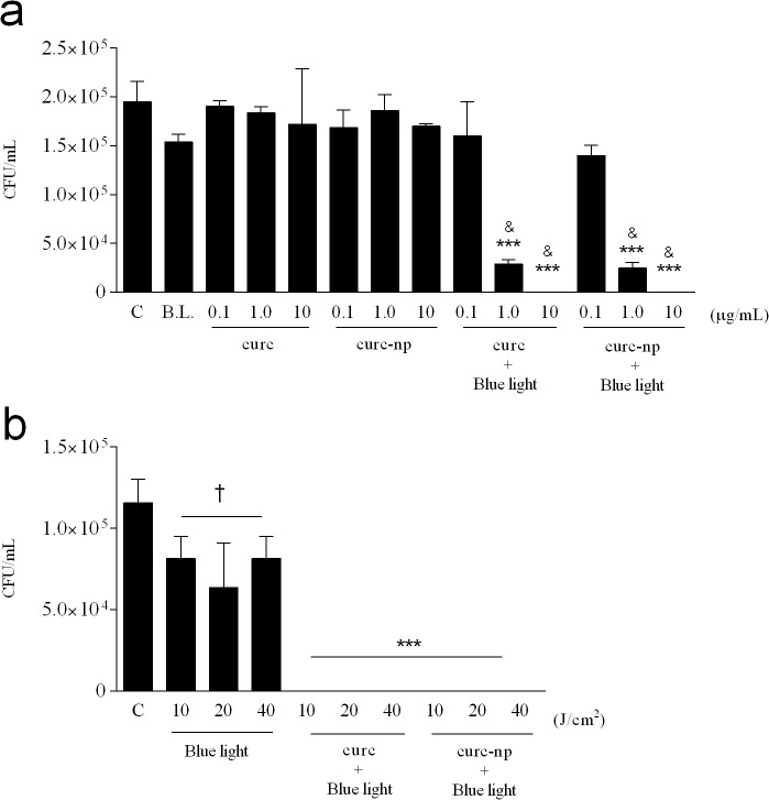
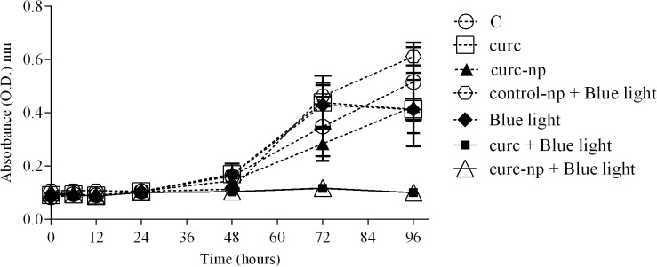
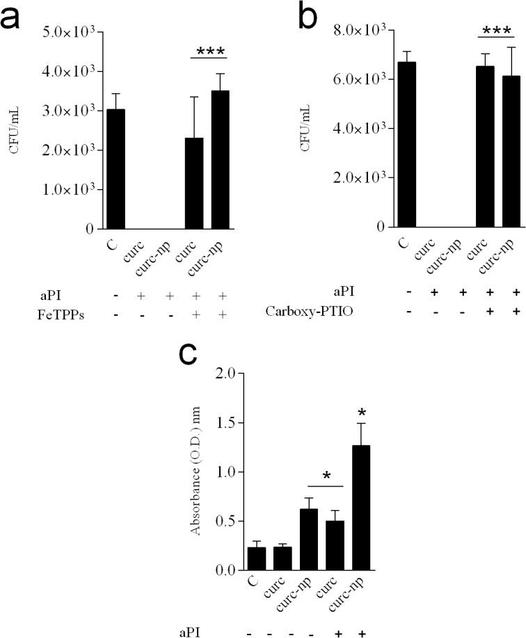
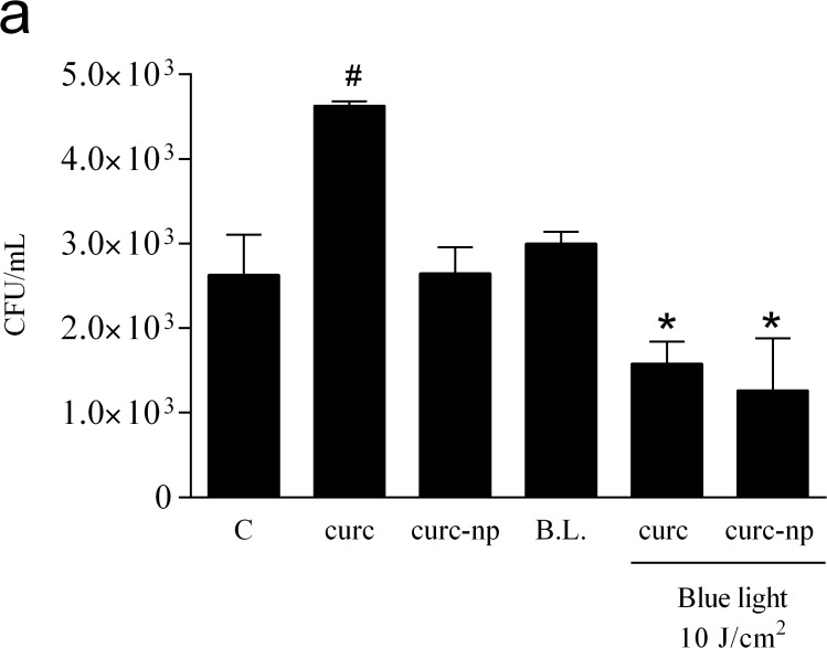
Antimicrobial photodynamic inhibition (aPI) utilizes radical stress generated from the excitation of a photosensitizer (PS) with light to destroy pathogens. Its use against Trichophyton rubrum, a dermatophytic fungus with increasing incidence and resistance, has not been well characterized. Our aim was to evaluate the mechanism of action of aPI against T. rubrum using curcumin as the PS in both free and nanoparticle (curc-np) form. Nanocarriers stabilize curcumin and allow for enhanced solubility and PS delivery. Curcumin aPI, at optimal conditions of 10 μg/mL of PS with 10 J/cm² of blue light (417 ± 5 nm), completely inhibited fungal growth (p<0.0001) via induction of reactive oxygen (ROS) and nitrogen species (RNS), which was associated with fungal death by apoptosis. Interestingly, only scavengers of RNS impeded aPI efficacy, suggesting that curcumin acts potently via a nitrosative pathway. The curc-np induced greater NO˙ expression and enhanced apoptosis of fungal cells, highlighting curc-np aPI as a potential treatment for T. rubrum skin infections.


Conflict of interest statement

Competing Interests: The authors have declared that no competing interests exist.

Figures

Fig 1
Fig 1. Optimization of aPI conditions.
(a) Effect of varying the PS concentration on fungal growth, as determined by colony forming units (CFU), using a constant light source of 40 J/cm2. (b) Effect of varying the light dose using a constant PS concentration of 10 μg/mL. Untreated T. rubrum (C), Blue light alone (B.L.) and PS without photoactivation (curc and curc-np) were used as controls. ***Compared to untreated, blue light and PS without photoactivation. &Compared to lowest PS concentration of same group. Compared to untreated control. ***p < 0.0001, &, †p < 0.05. Data are a composite of three independent experiments with each treatment group performed in triplicate. The results are expressed as the mean ± SEM.
Fig 2
Fig 2. Fungal growth curve after aPI.
Fungal growth curve of aPI at optimal conditions (10 μg/mL of PS with 10 J/cm2 of B.L.). Each treatment per group was performed in triplicate and data are a composite of three independent experiments. The results are expressed as the mean ± SEM.
Fig 3
Fig 3. Evaluation of ROS and RNS production after aPI.
Detection of ROS levels following aPI, expressed as a (a) representative histogram and (d) cumulative bar plot. Detection of NO levels following aPI, expressed as a (b) representative histogram and (e) cumulative bar plot. Detection of ONOO levels following aPI, expressed as a (c) representative histogram and (f) cumulative bar plot. Dark toxicity controls did not differ significantly from untreated T. rubrum (data not represented). ***Compared to untreated control. ###Compared to curc group. MFI. Mean fluorescence intensity. ***,###p < 0.0001. Each treatment per group was performed in triplicate and are a composite of two independent experiments. The results are expressed as the mean ± SEM.
Fig 4
Fig 4. Evaluation of aPI mechanism of action.
(a) Treatment with ONOO scavenger (FeTPPs). (b) Treatment with NO scavenger (Carboxy-PTIO). (c) Apoptosis assay performed after aPI. ***Compared to aPI treatment in the absence of incubation with scavengers. *Compared to untreated T. rubrum control. *p< 0.05, ***p< 0.0001. Each treatment per group was performed in triplicate and data are a composite of two independent experiments. The results are expressed as mean ± SEM.
Fig 5
Fig 5. Phagocytosis assay.
CFU quantification of macrophages challenged with T. rubrum cells and treated with aPI therapy. # Compared to untreated control (C), dark toxicity and blue light 10 J/cm2 (B.L.) controls. * Compared to all other groups. B.L. Blue light 10 J/cm2 (17 minutes). *,# p < 0.05. Each treatment per group was performed in triplicate and data is a composite of two independent experiments. The results are expressed as the mean ± SEM.

References

    1. Vermout S, Tabart J, Baldo A, Mathy A, Losson B, Mignon B. Pathogenesis of dermatophytosis. Mycopathologia. 2008; 166: 267–275. 10.1007/s11046-008-9104-5 - DOI - PubMed
    1. Gupta AK, Humke S. The prevalence and management of onychomycosis in diabetic patients. Eur J Dermatol. 2000; 10: 379–384. - PubMed
    1. Manzano-Gayosso P, Mendez-Tovar LJ, Hernandez-Hernandez F, Lopez-Martinez R [Antifungal resistance: an emerging problem in Mexico]. Gac Med Mex. 2008; 144: 23–26. - PubMed
    1. Martinez-Rossi NM, Peres NT, Rossi A. Antifungal resistance mechanisms in dermatophytes. Mycopathologia. 2008; 166: 369–383. 10.1007/s11046-008-9110-7 - DOI - PubMed
    1. Ghelardi E, Celandroni F, Gueye SA, Salvetti S, Senesi S, Bulgheroni A, et al. Potential of Ergosterol synthesis inhibitors to cause resistance or cross-resistance in Trichophyton rubrum . Antimicrob Agents Chemother. 2014; 58: 2825–2829. 10.1128/AAC.02382-13 - DOI - PMC - PubMed

Publication types

MeSH terms

LinkOut - more resources