. 2015 Mar 24;11(3):e1004687.
doi: 10.1371/journal.ppat.1004687. eCollection 2015 Mar.

β-HPV 5 and 8 E6 disrupt homology dependent double strand break repair by attenuating BRCA1 and BRCA2 expression and foci formation

Affiliations

β-HPV 5 and 8 E6 disrupt homology dependent double strand break repair by attenuating BRCA1 and BRCA2 expression and foci formation

Nicholas A Wallace et al. PLoS Pathog. .

Abstract

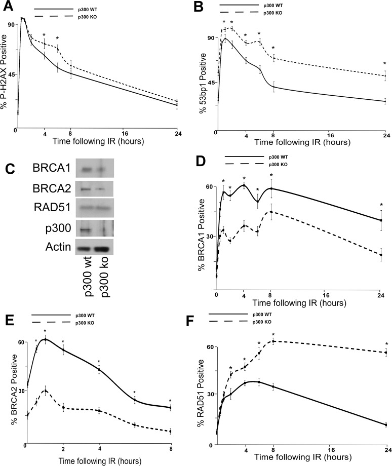
Recent work has explored a putative role for the E6 protein from some β-human papillomavirus genus (β-HPVs) in the development of non-melanoma skin cancers, specifically β-HPV 5 and 8 E6. Because these viruses are not required for tumor maintenance, they are hypothesized to act as co-factors that enhance the mutagenic capacity of UV-exposure by disrupting the repair of the resulting DNA damage. Supporting this proposal, we have previously demonstrated that UV damage signaling is hindered by β-HPV 5 and 8 E6 resulting in an increase in both thymine dimers and UV-induced double strand breaks (DSBs). Here we show that β-HPV 5 and 8 E6 further disrupt the repair of these DSBs and provide a mechanism for this attenuation. By binding and destabilizing a histone acetyltransferase, p300, β-HPV 5 and 8 E6 reduce the enrichment of the transcription factor at the promoter of two genes critical to the homology dependent repair of DSBs (BRCA1 and BRCA2). The resulting diminished BRCA1/2 transcription not only leads to lower protein levels but also curtails the ability of these proteins to form repair foci at DSBs. Using a GFP-based reporter, we confirm that this reduced foci formation leads to significantly diminished homology dependent repair of DSBs. By deleting the p300 binding domain of β-HPV 8 E6, we demonstrate that the loss of robust repair is dependent on viral-mediated degradation of p300 and confirm this observation using a combination of p300 mutants that are β-HPV 8 E6 destabilization resistant and p300 knock-out cells. In conclusion, this work establishes an expanded ability of β-HPV 5 and 8 E6 to attenuate UV damage repair, thus adding further support to the hypothesis that β-HPV infections play a role in skin cancer development by increasing the oncogenic potential of UV exposure.


Conflict of interest statement

The authors have declared that no competing interests exist.

Figures

Fig 1
Fig 1. β-HPV 5 and 8 E6 attenuate double strand break repair.
HFK cells were exposed to 4 gray of ionizing radiation and immunofluorescence microscopy was used to determine the percentage of cells that were positive for either (A.) p-H2AX foci or (B.) 53bp1 foci over a 24 hour period following irradiation. n ≥ 5. (C.) Representative images of cells stained for p-H2AX (pink) and nuclei (blue) 6 hours after exposure to IR. (D.) Representative images of cells stained for 53bp1 (pink) and nuclei (blue) 24 hours after exposure to IR. (E.) Chart depicts the mean number of breaks per chromosome measured in control cells as well as cells exposed to a DNA damaging agent. n = 3. Error bars depict the standard error of the mean. * denotes a significant difference of both β-HPV 5 and 8 E6 from vector control (LXSN) cells as determined by student t-test with a p value ≤ 0.05. # denotes that β-HPV Δ8 E6 was significantly different from corresponding wt β-HPV 8 E6 with a p-value ≤ 0.05 from a student T-test.
Fig 2
Fig 2. β-HPV 5 and 8 E6 attenuate HDR protein activation, abundance and resolution of damage-induced nuclear foci.
(A.) Representative immunoblot showing BRCA1, BRCA2, RPA70 and RAD51 protein levels in HFK cells. Nucleolin is shown as a loading control. (B.) Densitometry of immunoblots (n = 3) of BRCA1, BRCA2, RPA70 and RAD51. The amount of protein in each sample was normalized to the corresponding amount of nucleolin and then data was set relative to vector control (LXSN) cells. L denotes vector control (LXSN) HFKs. 5 denotes HPV 5 E6 expressing HFKs. 8 denotes HPV 8 E6 expressing HFKs. (D.)-(G.) HFK cells were exposed to 4 gray of ionizing radiation and immunofluorescence microscopy was used to determine the percentage of cells positive for (C.) BRCA1, (D.) BRCA 2, (E.) RPA70 and (F.) RAD51 foci over a 6–24 hour period. Error bars represent standard errors of the mean. * denotes that both HPV 5 and 8 E6 are significantly different from vector control cells as determined by student T-test (p value ≤ 0.05). # denotes that β-HPV Δ8 E6 was significantly different from corresponding wt β-HPV 8 E6 with a p-value ≤ 0.05 from a student T-test.
Fig 3
Fig 3. β-HPV 5 and 8 E6 attenuate homology dependent repair of double strand breaks.
(A.) A representation of the DR-GFP cassette that is clonally integrated into U2OS cells. Briefly, the cassette consists of a promoter depicted here as a box with an arrow pointing to the right. Further, it contains two copies of a GFP gene (box labeled GFP). The first is a full copy of the gene interrupted by the recognition site for the rare cutting endonuclease, I-SceI, that is flanked by stop codons preventing read-through expression of GFP. The second copy of GFP is missing 5’ and 3’ sequence. Exogenous expression of I-SceI in these cells will introduce a double strand break in the first GFP gene that if repaired by homology dependent repair (labeled HDR) using the second gene as a template will result in a functional GFP gene. (B.) Immunofluorescence microscopy was used to measure p-H2AX foci persistence in U2OS DR-GFP cells following exposure to 4 gray of ionizing radiation. This chart depicts the percentage of p-H2AX positive cells 10 minutes and 6 hours after irradiation. (C.) This chart depicts the relative amount of homology dependent repair as measured by GFP positive cells following exogenous expression of I-SceI. Percentage of GFP positive β-HPV 5 and 8 E6 cells are given relative to the level of GFP positive vector control (LXSN) cells that were set to 100. (D.) Representative samples of FACS profiles used to generate data shown in (C.) Error Bars represent standard errors of the mean. n ≥ 3 for all experiments. * denotes significant difference from similarly treated control cells with a p-value ≤ 0.05 as determined by student T-test. # denotes that β-HPV Δ8 E6 was significantly different from corresponding wt β-HPV 8 E6 with a p-value ≤ 0.05 from a student T-test.
Fig 4
Fig 4. p300 is required for efficient double strand break repair.
HCT 116 cells either with p300 knocked out (p300 ko) or with those p300ko cells complimented with wt p300 (p300 wt) were exposed to 4 gray of ionizing radiation and immunofluorescence microscopy was used to observe (A.) p-H2AX and (B.) 53bp1 foci formation for 24 hours after irradiation. (C.) Representative immunoblot of HCT 116 cells showing RAD51, BRCA2, BRCA1 and p300. Actin is included as a loading control. (D.)–(F.) HCT 116 were exposed to 4 gray of ionizing radiation and immunofluorescence microscopy was used to observe BRCA1, BRCA2 and RAD51 foci formation for 8 or 24 hours after irradiation. For all experiments, error bars depict standard errors of the mean. Similarly, * denote points where HCT 116 p300 ko cells significantly differed from control cells by student T-test with a p value ≤ 0.05. Finally, all experiments were conducted at least three times independently.
Fig 5
Fig 5. A mutant p300 that is resistant to β-HPV E6 mediated degradation rescues homology dependent repair of DSBs.
Immunofluorescence microscopy was used to measure the intensity of (A.) BRCA1 and (B.) BRCA2 as well as foci positive cells for (C.) p-H2AX, (D.) BRCA1, (E.) BRCA2 and (F.) RAD51 foci persistence in HFK cells transfected with p300 expression vectors following exposure to 4 gray of ionizing radiation. These charts depict the percentage of foci positive cells at the indicated time points after irradiation. p300A denotes p300 S1834A mutant. p300E denotes p300 S1834E mutant. For all experiments, error bars depict standard errors of the mean. Similarly, * denote points where β-HPV E6 expressing cells significantly differed from the values of the corresponding control cells determined by student T-test and with a p value ≤ 0.05. # denotes a statistically significant difference within a cell line between cells transfected with p300A and p300E. Finally, all experiments were conducted at least three times independently.
Fig 6
Fig 6. β-HPV 5 and 8 E6 increases sensitivity to ionizing radiation and PARP1 inhibition.
(A.)–(C.) Relative viability of HFK cells exposed to either (A.) ionizing radiation (2 gray) or grown in media with PARP1 inhibitors (10 nM) (B.) AZD 2281 (AZD) and (C.) ABT 888 (ABT). n ≥ 3. Error bars represent standard errors of the mean. * denotes data that is statistically different from the similarly treated vector control as determined by student T-test with a p value ≤ 0.05. # denotes data that the value for HPV Δ8 E6 is statistically different from the similarly treated HPV 8 E6 cells as determined by student T-test with a p value ≤ 0.05. L denotes vector control (LXSN) HFKs. 5 denotes HPV 5 E6 expressing HFKs. 8 denotes HPV 8 E6 expressing HFKs. Δ8 denotes HPV Δ8 E6 expressing HFKs.

References

    1. Bernard HU, Burk RD, Chen Z, van Doorslaer K, Hausen H, et al. (2010) Classification of papillomaviruses (PVs) based on 189 PV types and proposal of taxonomic amendments. Virology 401: 70–79. 10.1016/j.virol.2010.02.002 - DOI - PMC - PubMed
    1. zur Hausen H (1999) Papillomaviruses in human cancers. Proc Assoc Am Physicians 111: 581–587. - PubMed
    1. zur Hausen H (2002) Papillomaviruses and cancer: from basic studies to clinical application. Nat Rev Cancer 2: 342–350. - PubMed
    1. Bouwes Bavinck JN, Feltkamp M, Struijk L, ter Schegget J (2001) Human papillomavirus infection and skin cancer risk in organ transplant recipients. J Investig Dermatol Symp Proc 6: 207–211. - PubMed
    1. Orth G, Jablonska S, Jarzabek-Chorzelska M, Obalek S, Rzesa G, et al. (1979) Characteristics of the lesions and risk of malignant conversion associated with the type of human papillomavirus involved in epidermodysplasia verruciformis. Cancer Res 39: 1074–1082. - PubMed

Publication types

MeSH terms

LinkOut - more resources