. 2015 Mar 24;10(3):e0121421.
doi: 10.1371/journal.pone.0121421. eCollection 2015.

CD105 expression on CD34-negative spindle-shaped stromal cells of primary tumor is an unfavorable prognostic marker in early breast cancer patients

Affiliations

CD105 expression on CD34-negative spindle-shaped stromal cells of primary tumor is an unfavorable prognostic marker in early breast cancer patients

Leandro Marcelo Martinez et al. PLoS One. .

Erratum in

Abstract

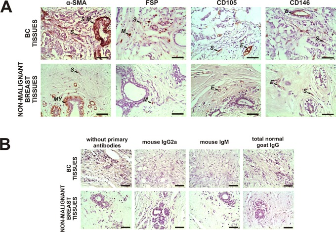
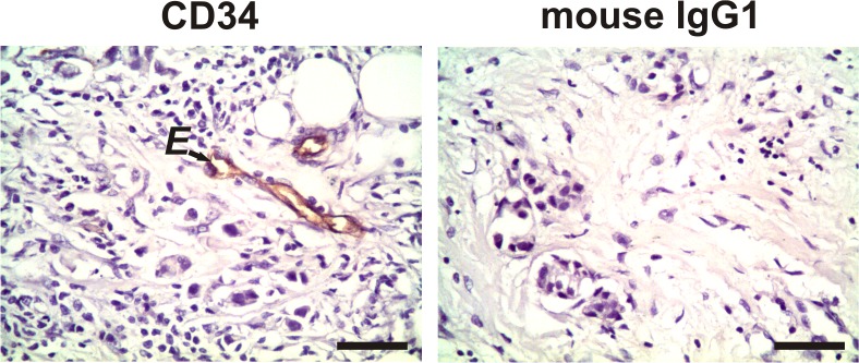
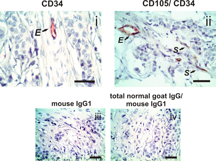
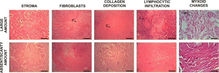
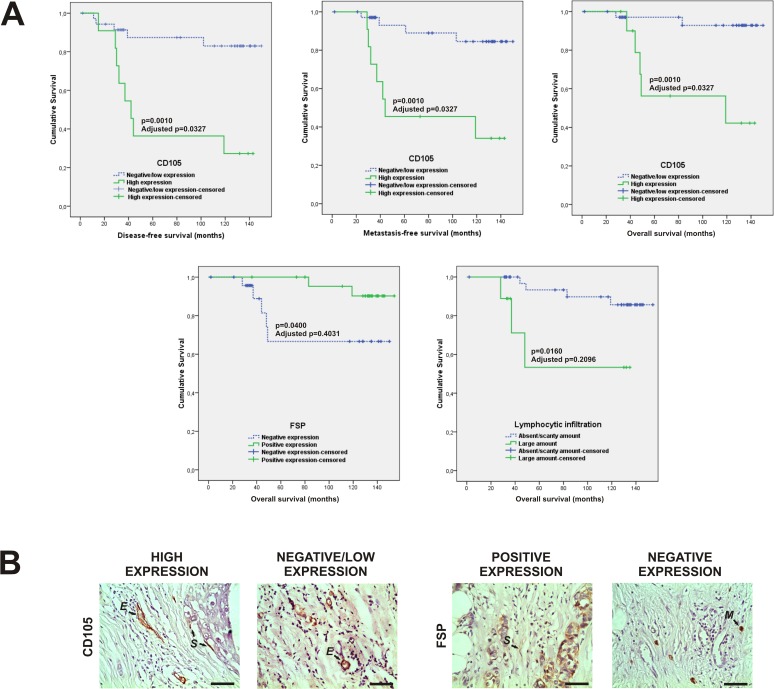
Several studies have confirmed that the breast tumor microenvironment drives cancer progression and metastatic development. The aim of our research was to investigate the prognostic significance of the breast tumor microenvironment in untreated early breast cancer patients. Therefore, we analyzed the association of the expression of α-SMA, FSP, CD105 and CD146 in CD34-negative spindle-shaped stromal cells, not associated with the vasculature, in primary breast tumors with classical prognostic marker levels, metastatic recurrence, local relapse, disease-free survival, metastasis-free survival and the overall survival of patients. In the same way, we evaluated the association of the amount of intra-tumor stroma, fibroblasts, collagen deposition, lymphocytic infiltration and myxoid changes in these samples with the clinical-pathological data previously described. This study is the first to demonstrate the high CD105 expression in this stromal cell type as a possible independent marker of unfavorable prognosis in early breast cancer patients. Our study suggests that this new finding can be useful prognostic marker in the clinical-pathological routine.


Conflict of interest statement

Competing Interests: The authors have declared that no competing interests exist.

Figures

Fig 1
Fig 1. Expression of α-SMA, FSP, CD105 and CD146 in malignant and non-malignant breast tissues.
A Representative immunohistochemistry staining for α-SMA, FSP, CD105 and CD146 in stromal cells of primary tumor tissues from breast cancer (BC) patients and non-malignant breast tissues. Reactions were evaluated in spindle-shaped stromal cells, not associated with the vasculature. The arrows show positive staining of evaluated stromal cells (S) and smooth muscle cells (S M), myoepithelial cells (MY), tissue-associated macrophages (M) and endothelial cells (E) used as internal positive controls as previously described. B No staining was observed in both types of tissues when we incubated them without primary antibodies and with irrelevant mouse IgG2a (for α-SMA), irrelevant mouse IgM (for FSP) or total normal goat IgG (for CD105 and CD146) as negative controls. Nuclei were counterstained with hematoxylin (purple). Original magnification A and B: 400×. The scale bars represent 50 μm.
Fig 2
Fig 2. Expression of CD34 in tumor stromal cells.
Representative negative staining for CD34 in the evaluated spindle-shaped stromal cells, not associated with the vasculature, of primary tumor tissue from a breast cancer patient. The arrow shows an example of CD34-positive staining only in the endothelial cells (E). No staining was observed in the tissues when we incubated them with irrelevant mouse IgG1 as a negative isotype control. Nuclei were counterstained with hematoxylin (purple). Original magnification: 400×. The scale bar represents 50 μm.
Fig 3
Fig 3. Expression of CD105 and CD34 in tumor stromal cells.
i Single immunohistochemistry for CD34 (detected by red chromogen) shows a representative example of CD34-positive staining in the endothelial cells (E). ii Double immunohistochemistry for CD105 and CD34 (detected by brown and red chromogen, respectively) shows a representative example of co-staining of CD105 and CD34 in the endothelial cells (E) and CD105-positive staining only in evaluated stromal cells (S) of primary tumor tissue from a breast cancer patient. No staining was observed in the tissues when we incubated them with irrelevant mouse IgG1 (for CD34) or sequentially incubated with total normal goat IgG and irrelevant mouse IgG1 (for CD105/ CD34) as negative isotype controls (iii and iv; respectively). Nuclei were counterstained with hematoxylin (purple). Original magnification: 400×. The scale bars represent 50 μm.
Fig 4
Fig 4. Histological features of primary breast tumor stroma as determined by hematoxylin and eosin staining.
This picture is an example of samples with a large amount and absent/scanty amount of stroma, fibroblasts, collagen deposition, lymphocytic infiltration and myxoid changes. The arrows show tumor fibroblasts (F), collagen deposition (C), lymphocytic infiltration (L) and myxoid changes (M C). Original magnification: 100×. The scale bars represent 200 μm.
Fig 5
Fig 5. Correlation of CD105 and FSP expression, as well as lymphocytic infiltration quantity with disease-free survival, metastasis-free survival and overall survival of 56 untreated early breast cancer patients.
A Kaplan-Meier curves show that high stromal cell expression of CD105 is associated with a shorter disease-free survival, metastasis-free survival and overall survival of our breast cancer patients. The difference is statistically significant [p = 0.0010 in all of cases by log-rank (Mantel-Cox)-test]. The adjusted p-value is 0.0327 (Benjamini-Hochberg correction) in all of cases. Moreover, positive FSP expression is significantly associated with a greater overall survival of these patients [p = 0.0400 by log-rank (Mantel-Cox) test]. Furthermore, a large amount of lymphocytic infiltration is significantly associated with a shorter overall survival of these patients [p = 0.0160, log-rank (Mantel-Cox) test]. After the Benjamini-Hochberg adjustment, these last associations were considered not significant (adjusted p = 0.4031 and 0.2096, respectively). B Photographs show a representative immunohistochemistry staining for tumor samples with high and negative/low stromal expression of CD105, as well as tumor samples with positive and negative FSP expression. The arrows show positive staining of evaluated stromal cells (S), endothelial cells (E) and tissue associated-macrophages (M) used as internal positive controls as previously described. Original magnification: 400×. The scale bars represent 50 μm.

References

    1. Patel SA, Heinrich AC, Reddy BY, Srinivas B, Heidaran N, Rameshwar P. Breast cancer biology: the multifaceted roles of mesenchymal stem cells. J Oncol. 2008; 2008: 425895 10.1155/2008/425895 - DOI - PMC - PubMed
    1. Mao Y, Keller ET, Garfield DH, Shen K, Wang J. Stromal cells in tumor microenvironment and breast cancer. Cancer Metastasis Rev. 2013; 32: 303–315. 10.1007/s10555-012-9415-3 - DOI - PMC - PubMed
    1. Paulsson J, Micke P. Prognostic relevance of cancer-associated fibroblasts in human cancer. Semin Cancer Biol. 2014; 25: 61–68. 10.1016/j.semcancer.2014.02.006 - DOI - PubMed
    1. Khamis ZI, Sahab ZJ, Sang QX. Active roles of tumor stroma in breast cancer metastasis. Int J Breast Cancer. 2012; 2012: 574025 10.1155/2012/574025 - DOI - PMC - PubMed
    1. Joyce JA, Pollard JW. Microenvironmental regulation of metastasis. Nat Rev Cancer. 2009; 9: 239–252. 10.1038/nrc2618 - DOI - PMC - PubMed

Publication types

MeSH terms