Case Reports
. 2015 Mar 24;10(3):e0120841.
doi: 10.1371/journal.pone.0120841. eCollection 2015.

Screening of drugs to treat 8p11 myeloproliferative syndrome using patient-derived induced pluripotent stem cells with fusion gene CEP110-FGFR1

Affiliations
Case Reports

Screening of drugs to treat 8p11 myeloproliferative syndrome using patient-derived induced pluripotent stem cells with fusion gene CEP110-FGFR1

Shohei Yamamoto et al. PLoS One. .

Abstract

Induced pluripotent stem (iPS) cells provide powerful tools for studying disease mechanisms and developing therapies for diseases. The 8p11 myeloproliferative syndrome (EMS) is an aggressive chronic myeloproliferative disorder (MPD) that is caused by constitutive activation of fibroblast growth factor receptor 1. EMS is rare and, consequently, effective treatment for this disease has not been established. Here, iPS cells were generated from an EMS patient (EMS-iPS cells) to assist the development of effective therapies for EMS. When iPS cells were co-cultured with murine embryonic stromal cells, EMS-iPS cells produced more hematopoietic progenitor and hematopoietic cells, and CD34+ cells derived from EMS-iPS cells exhibited 3.2-7.2-fold more macrophage and erythroid colony forming units (CFUs) than those derived from control iPS cells. These data indicate that EMS-iPS cells have an increased hematopoietic differentiation capacity, which is characteristic of MPDs. To determine whether a tyrosine kinase inhibitor (TKI) could suppress the increased number of CFUs formed by EMS-iPS-induced CD34+ cells, cells were treated with one of four TKIs (CHIR258, PKC 412, ponatinib, and imatinib). CHIR258, PKC 412, and ponatinib reduced the number of CFUs formed by EMS-iPS-induced CD34+ cells in a dose-dependent manner, whereas imatinib did not. Similar effects were observed on primary peripheral blood cells (more than 90% of which were blasts) isolated from the patient. This study provides evidence that the EMS-iPS cell line is a useful tool for the screening of drugs to treat EMS and to investigate the mechanism underlying this disease.


Conflict of interest statement

Competing Interests: The authors have declared that no competing interests exist.

Figures

Fig 1
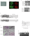
Fig 1. Generation of iPS cells from an EMS patient with the t(8;9)(p12;q33) translocation.
(a) Morphology of EMS-iPS cells (×40 magnification). (b) Expression of the pluripotency markers Nanog (A), SOX2 (B), OCT3/4 (C), and PODXL (D) in EMS-iPS cells (×10 magnification). (c) Semi-quantitative RT-PCR analysis of the expression levels of endogenous (end) and viral-derived (Tg) reprogramming factors. Primary BM cells from the patient and control 201B7 human iPS cells (B7) are included as controls. (d) Bisulfite sequencing analyses of the OCT3/4 and NANOG promoter regions in patient BM cells and EMS-iPS cells. White and black circles represent unmethylated and methylated (Me) CpG dinucleotides, respectively. (e) Hematoxylin and eosin staining of a teratoma derived from EMS-iPS cells (×20 magnification). The teratoma is composed of gut-like epithelium (endoderm), skeletal muscle (mesoderm), and melanocytes (ectoderm). (f) EMS-iPS cells exhibit the 46, XY, t(8;9)(p12;q33) karyotype, as determined by G-banding analysis. (g) PCR analysis of the expression level of the CEP110-FGFR1 fusion transcript in primary BM cells from the patient and EMS-iPS cells.
Fig 2
Fig 2. EMS-iPS cells exhibit augmented hematopoiesis.
(a) Cobblestone morphology of iPS cells. Control human 201B7 iPS cells (B7) or EMS-iPS cells were co-cultured with murine AGM-S3 cells. After 12 days of co-culture, cells with a cobblestone morphology were detected at the peripheries of colonies. Images on the left are at ×100 magnification. Images on the right show higher magnification (×200 magnification) images of the boxed regions. (b) Flow cytometric analysis of cells cocultured with AGMS-3 on day 12. Cells were stained with antibodies specific CD34 and CD45. The percentages of CD34+ and /or CD45+ cells were shown (n = 3; bars represent SDs, p<0.05).
Fig 3
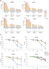
Fig 3. Effects of TKIs on hematopoiesis of EMS-iPS cells.
(a) EMS-iPS cells and control iPS cells (control 1: 201B7 and control 2: TkDA3-1) generated by expression of four factors were co-cultured with AGM-S3 cells. After 12 days, cells derived from iPS cells were collected. CD34+ cells were isolated for clonal hematopoietic culture in the presence of various concentrations of CHIR258, PKC412, ponatinib, or imatinib. Data show mean number of colonies formed ± SD. *P < 0.05 compared to cells cultured in the absence of TKIs. (b) EMS-iPS cells generated by expression of four factors were co-cultured with AGM-S3 cells. After 12 days, cells derived from iPS cells were collected. CD34+ cells were isolated for suspension culture in the absence or presence of various concentrations of CHIR258, PKC412, ponatinib, or imatinib. Data show the mean ± SD of the percentage of viable cells compared to the number in untreated samples, which was set at 100%. *P < 0.05 compared to cells cultured in the absence of TKIs.
Fig 4
Fig 4. Effect of TKIs on primary PB cells of the EMS patient.
(a) May-Grünwald Giemsa staining of a cytospin preparation of primary PB cells from the EMS patient (×100 magnification). (b) PM MNCs (more than 90% of which were blasts) from the EMS patient were cultured in suspension in the presence of CHIR258, PKC412, ponatinib, or imatinib for 7 days. Data show mean ± SD of the percentage of viable cells compared to the number in untreated samples, which was set at 100%. *P < 0.05 compared to cells cultured in the absence of TKIs (control).

References

    1. Macdonald D, Aguiar RC, Mason PJ, Goldman JM, Cross NC. A new myeloproliferative disorder associated with chromosomal translocations involving 8p11: a review. Leukemia. 1995;9(10):1628–30. Epub 1995/10/01. PubMed . - PubMed
    1. Macdonald D, Reiter A, Cross NC. The 8p11 myeloproliferative syndrome: a distinct clinical entity caused by constitutive activation of FGFR1. Acta haematologica. 2002;107(2):101–7. Epub 2002/03/29. doi: 46639. PubMed . - PubMed
    1. Jackson CC, Medeiros LJ, Miranda RN. 8p11 myeloproliferative syndrome: a review. Human pathology. 2010;41(4):461–76. Epub 2010/03/17. 10.1016/j.humpath.2009.11.003 PubMed . - DOI - PubMed
    1. Hu S, He Y, Zhu X, Li J, He H. Myeloproliferative disorders with t(8;9)(p12;q33): a case report and review of the literature. Pediatric hematology and oncology. 2011;28(2):140–6. Epub 2011/01/11. 10.3109/08880018.2010.528170 PubMed . - DOI - PubMed
    1. Guasch G, Mack GJ, Popovici C, Dastugue N, Birnbaum D, Rattner JB, et al. FGFR1 is fused to the centrosome-associated protein CEP110 in the 8p12 stem cell myeloproliferative disorder with t(8;9)(p12;q33). Blood. 2000;95(5):1788–96. Epub 2000/02/26. PubMed . - PubMed

Publication types

MeSH terms

LinkOut - more resources