Review
. 2015 Dec;23(12):1634-9.
doi: 10.1038/ejhg.2015.41. Epub 2015 Mar 25.

The lymphatic phenotype in Turner syndrome: an evaluation of nineteen patients and literature review

Affiliations
Review

The lymphatic phenotype in Turner syndrome: an evaluation of nineteen patients and literature review

Giles Atton et al. Eur J Hum Genet. 2015 Dec.

Abstract

Turner syndrome is a complex disorder caused by an absent or abnormal sex chromosome. It affects 1/2000-1/3000 live-born females. Congenital lymphoedema of the hands, feet and neck region (present in over 60% of patients) is a common and key diagnostic indicator, although is poorly described in the literature. The aim of this study was to analyse the medical records of a cohort of 19 Turner syndrome patients attending three specialist primary lymphoedema clinics, to elucidate the key features of the lymphatic phenotype and provide vital insights into its diagnosis, natural history and management. The majority of patients presented at birth with four-limb lymphoedema, which often resolved in early childhood, but frequently recurred in later life. The swelling was confined to the legs and hands with no facial or genital swelling. There was only one case of suspected systemic involvement (intestinal lymphangiectasia). The lymphoscintigraphy results suggest that the lymphatic phenotype of Turner syndrome may be due to a failure of initial lymphatic (capillary) function.


Figures

Figure 1
Figure 1
Clinical images of patient 7 (a(i–iv)) and patient 6 (b(i–iv)). (a(i), b(i)) Anterior and lateral view of the head and neck demonstrating some of the following features of Turner syndrome: a webbed neck, a low posterior hairline, low-set posteriorly rotated ears and a broad chest. (a(ii), b(ii)) Hands: Bilateral swelling of the hands and fingers. (a(iii), b(iii)) Below-knee: Bilateral swelling of the legs and feet. a(iv) Toes: Swelling with papillomatosis, small dysplastic toenails and deep digital skin fold creases. B(iv) Toes: Swelling with small dysplastic toenails.
Figure 2
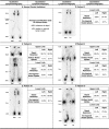
Figure 2
Anteroposterior lymphoscintigrams at 2 h following an interstitial injection of a radiolabelled colloid tracer (Technetium (99mTc)). (a) Normal control individual: Lower limb lymphoscintigram. Patent lymphatic channels and inguinal nodes imaged. Average quantification data for normal limbs demonstrate <80% retention at depot and >8% uptake in regional lymph nodes. (b) Patient 2, lower limb lymphoscintigram: Symmetrical drainage to inguinal lymph nodes with reduced imaging of the lymphatic channels bilaterally. Quantitative lymphoscintigraphy demonstrates a significant bilateral retention of tracer in the feet after 2 h of 100% (left) and 97.4% (right). (c) Patient 6, four-limb limb lymphoscintigram: Upper limbs: Quantitative lymphoscintigraphy demonstrates a retention of tracer in the hands after 2 h of 96.1% (left) and 92.3% (right) with a concurrent markedly reduced axillary uptake of 1.1% (left) and 2.7% (right). Lower limb: Quantitative lymphoscintigraphy demonstrates a retention of tracer in the left foot (95.2%) but not the right (72.7%). Bilateral lower limb swelling that is worse on the left is commensurate with these lymphoscintigraphy findings. (d) Patient 8, four-limb lymphoscintigram: Upper limbs: Quantitative lymphoscintigraphy demonstrates a retention of tracer in the hands after 2 h of 93.6% (left) and 92.5% (right). Lower limb: Evidence of rerouting in the right lower limb. Quantitative lymphoscintigraphy demonstrates that the percentage retention of tracer in the feet after 2 h is 80.1% (left) and 82.5% (right). Abnormalities are observed in the imaged drainage routes rather than drainage function. (e) Patient 18, lower limb lymphoscintigram: Bilateral drainage to the inguinal lymph nodes. Reduced imaging of lymphatic channels on the left. Imaging of popliteal lymph nodes on the left suggests deep rerouting. Quantitative lymphoscintigraphy demonstrates an adequate percentage drainage of tracer to the ilioinguinal lymph nodes after 2 h of 14.3% (left) and 9.1% (right). Reduced drainage on the right is consistent with clinical lymphoedema of the right lower limb. (f) Patient 19, four-limb lymphoscintigram: Upper limb: Quantitative lymphoscintigraphy demonstrates a retention of tracer in the hands after 2 h of 93.9% (left) and 92.6% (right). Lower limb: Asymmetrical drainage to inguinal lymph nodes with no main lymphatic tracts imaged and significant dermal rerouting. Quantitative lymphoscintigraphy determined that the percentage retention of tracer in the feet after 2 h is 91.4% (left) and 77.8% (right). Ilioinguinal nodal uptake was markedly reduced at 1.3% (left) and 0.1% (right).

References

    1. Wolff DJ, Van Dyke DL, Powell CM: Working Group of the ACMG Laboratory Quality Assurance Committee Laboratory guideline for Turner syndrome. Genet Med 2010; 12: 52–55. - PubMed
    1. Gunther DF, Sybert VP: Lymphatic, tooth and skin manifestations in Turner syndrome. Int Congr Ser 2006; 1298: 58–62.
    1. Zhong Q, Layman LC: Genetic considerations in the patient with Turner syndrome - 45,X with or without mosaicism. Fertil Steril 2012; 98: 775–779. - PMC - PubMed
    1. Matura LA, Ho VB, Rosing DR, Bondy CA: Aortic dilatation and dissection in Turner syndrome. Circulation 2007; 116: 1663–1670. - PubMed
    1. Sybert VP, McCauley E: Turner's Syndrome. N Engl J Med 2004; 351: 1227–1238. - PubMed