. 2015 Apr;27(4):984-1001.
doi: 10.1105/tpc.15.00010. Epub 2015 Mar 24.

Spontaneous dominant mutations in chlamydomonas highlight ongoing evolution by gene diversification

Affiliations

Spontaneous dominant mutations in chlamydomonas highlight ongoing evolution by gene diversification

Alix Boulouis et al. Plant Cell. 2015 Apr.

Abstract

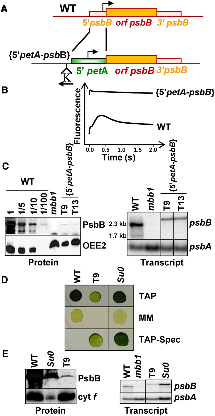
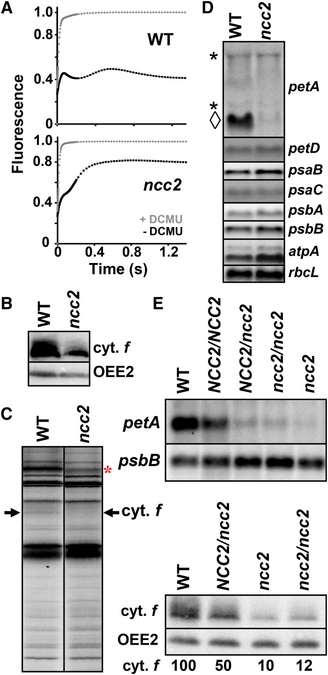
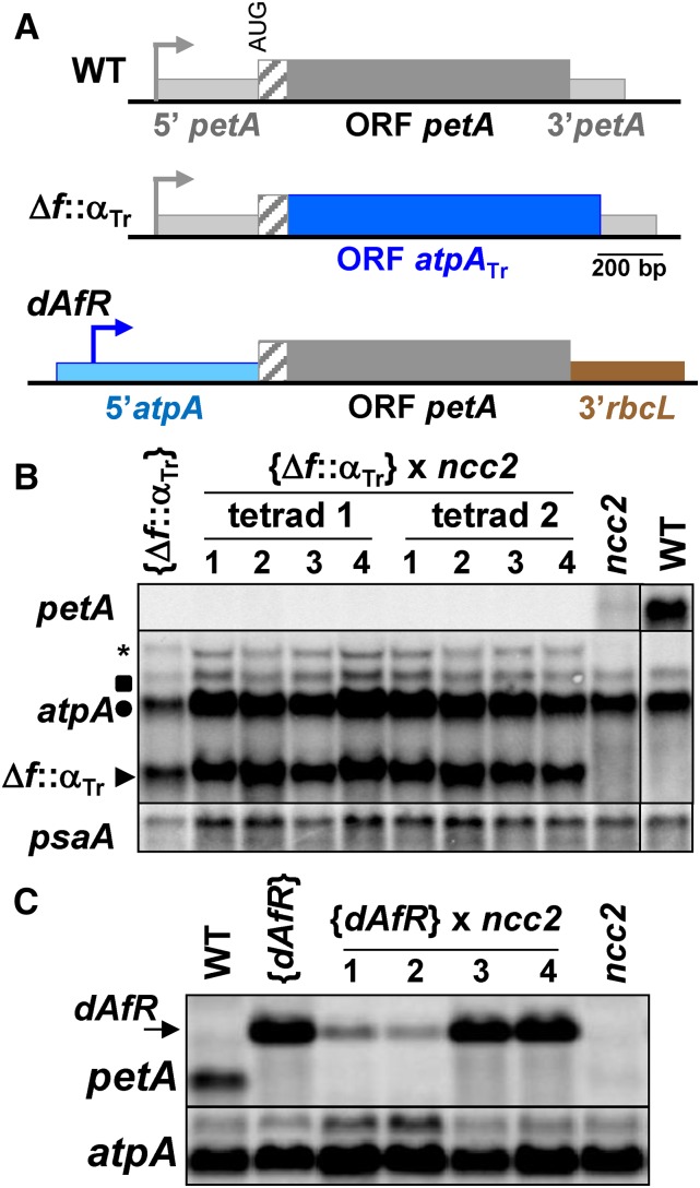
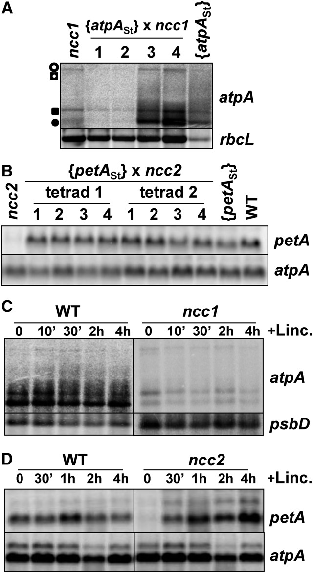
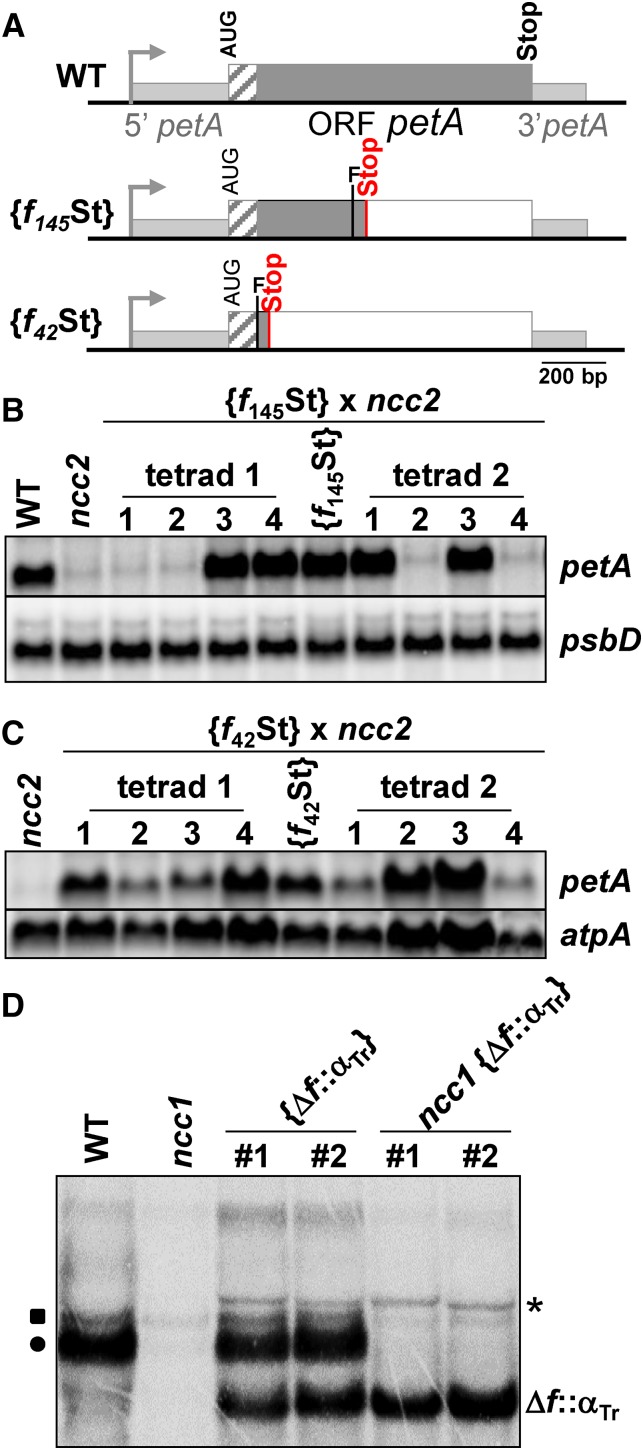
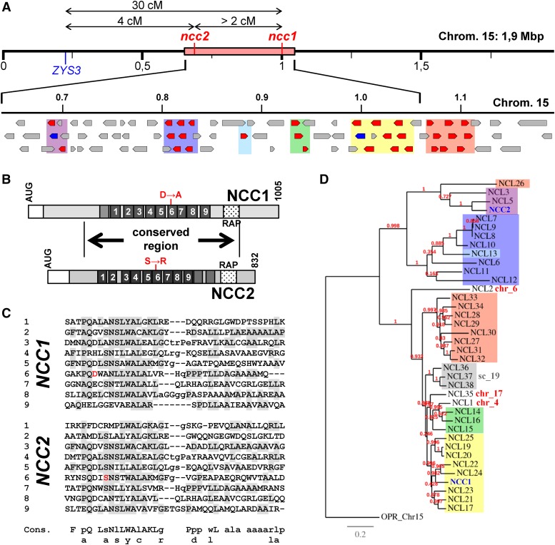
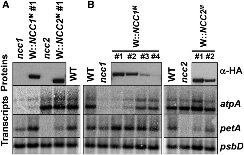
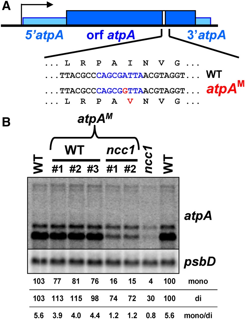
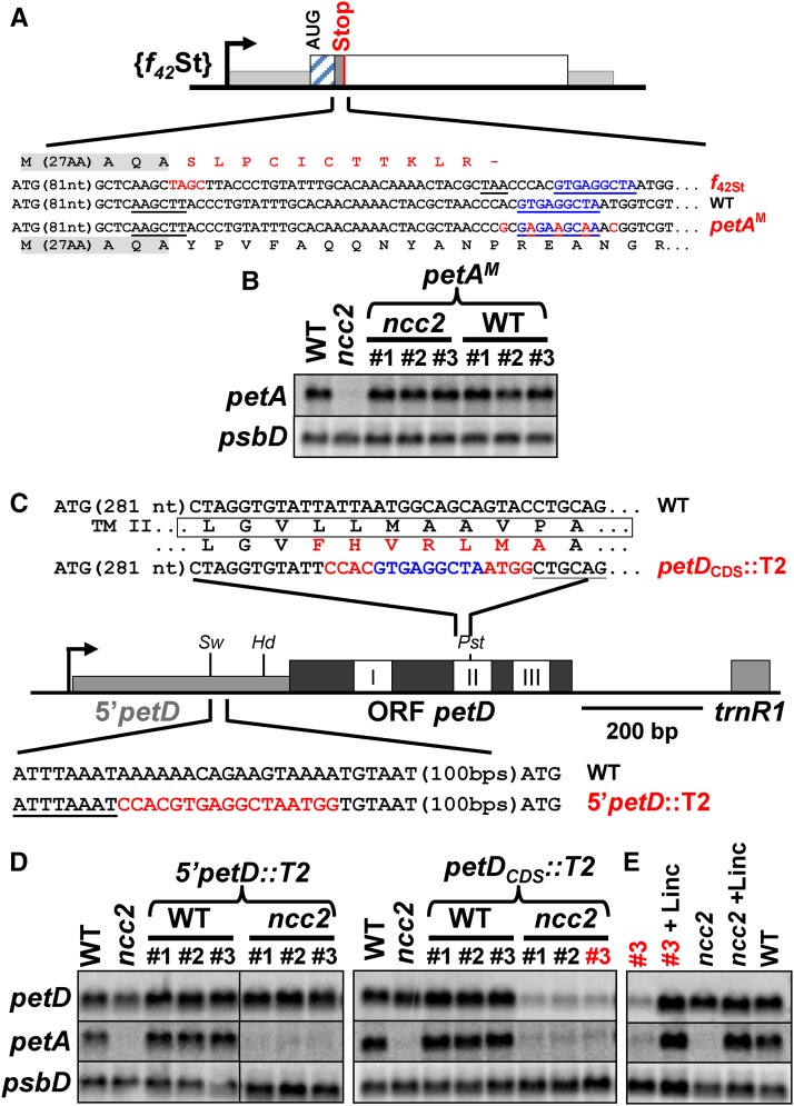
We characterized two spontaneous and dominant nuclear mutations in the unicellular alga Chlamydomonas reinhardtii, ncc1 and ncc2 (for nuclear control of chloroplast gene expression), which affect two octotricopeptide repeat (OPR) proteins encoded in a cluster of paralogous genes on chromosome 15. Both mutations cause a single amino acid substitution in one OPR repeat. As a result, the mutated NCC1 and NCC2 proteins now recognize new targets that we identified in the coding sequences of the chloroplast atpA and petA genes, respectively. Interaction of the mutated proteins with these targets leads to transcript degradation; however, in contrast to the ncc1 mutation, the ncc2 mutation requires on-going translation to promote the decay of the petA mRNA. Thus, these mutants reveal a mechanism by which nuclear factors act on chloroplast mRNAs in Chlamydomonas. They illustrate how diversifying selection can allow cells to adapt the nuclear control of organelle gene expression to environmental changes. We discuss these data in the wider context of the evolution of regulation by helical repeat proteins.


Figures

Figure 1.
Figure 1.
Isolation of the ncc2 Mutant. (A) Schematic description of the psbB gene in wild-type and {5′petA-psbB} transformants. Bent arrows indicate promoters, and the position and orientation of the selection cassette (K) are indicated. (B) Fluorescence induction kinetics of dark-adapted wild-type and {5′petA-psbB} cells. The nearly constant fluorescence intensity over time in strain {5′petA-psbB}, as well as its high initial level, almost similar to the stationary level, is typical of leaky PSII mutants. (C) Phenotypic characterization of {5′petA-psbB} transformed strains. Left: PsbB accumulation probed with a specific antibody in two independent transformants, in a dilution series of the wild type and in the mbb1-222E strain, defective for the accumulation of the psbB mRNA (Monod et al., 1992), as a specificity control. OEE2, whose accumulation is independent of PSII assembly (de Vitry et al., 1989), provides a loading control. Right: psbB mRNA accumulation in the same strains. Because of the larger size of the petA versus psbB 5′UTRs, the chimeric mRNA migrates more slowly than the endogenous psbB transcript. (D) Growth properties of the wild-type, {5′petA-psbB}, and Su0 strains. Drops of liquid culture (2 × 10⁶ cells⋅mL−1) were spotted on TAP medium and grown under dim light (10 μE⋅m−2⋅s−1, top), on minimal medium (MM) under high light (150 μE⋅m−2⋅s−1, intermediate), or on TAP supplemented with spectinomycin (500 μg⋅mL−1; lower panel). Pictures were taken after 10 d of growth. (E) Phenotypic characterization of the Su0 strain. Accumulation of PsbB and cytochrome f (left) and of the psbB mRNA (right) in the same strains. psbA was used as a loading control.
Figure 2.
Figure 2.
The Dominant ncc2 Mutation Confers a b6f Leaky Phenotype Due to Reduced Accumulation of the petA mRNA. (A) Fluorescence induction kinetics of dark-adapted ncc2 and wild-type cells. Black and gray curves show the kinetics recorded in the absence and in the presence, respectively, of DCMU (5 μM), which blocks electron transfer downstream of PSII. Maximal fluorescence levels in the presence of DCMU were normalized to 1. (B) Cytochrome f accumulation in wild-type and ncc2 strains. OEE2 provides a loading control. (C) Translation of chloroplast genes determined by 5 min 14C-acetate (5 μCi⋅mL−1) pulse-labeling experiments performed in the presence of cycloheximide (10 μg⋅mL−1) to block cytosolic translation. The arrow indicates the position of cytochrome f. The origin of the reduced rate of synthesis of Rubisco LS (red asterisk) in the ncc2 mutant, despite the unchanged accumulation of the rbcL transcript (D), is unknown. (D) Accumulation of representative transcripts for chloroplast photosynthesis genes in wild-type and ncc2 strains, assessed by RNA gel blots using the probes indicated on the right of the panel. For the petA gene, the diamond indicates the mature transcript, while asterisks point to precursor RNA species. (E) Accumulation of petA mRNA and cytochrome f, determined as above, in diploid strains either homozygous or heterozygous for the ncc2 mutation. Wild-type and ncc2 strains are shown for comparison. psbB mRNA and OEE2 provide loading controls in RNA and immunoblots, respectively.
Figure 3.
Figure 3.
The ncc2 Mutation Targets the petA CDS. (A) Scheme of the chimeras. Sequence encoding the mature cytochrome f is shown as a dark-gray rectangle and that encoding the lumen targeting peptide as a hatched rectangle. petA 5′ and 3′UTRs are represented by thinner light-gray rectangles. petA promoter is indicated by a bent arrow. In Δf::αTr chimera, the region encoding mature cytochrome f was replaced by the first 944 nucleotides of the atpA CDS, depicted as a blue rectangle. In dAfR chimera, the petA promoter and 5′UTR regions were replaced by the corresponding atpA regions (pale-blue rectangle), while petA 3′UTR was replaced by that of the rbcL gene (brown rectangle). (B) Transcript accumulation in tetrad progeny from the cross {Δf::αTr} × ncc2 and in wild-type and parental strains. RNA gel blots were hybridized with probes derived from petA, atpA, and psaA (loading control) CDSs, as indicated on the left. Positions of the endogenous mono- and dicistronic atpA transcripts are indicated by a circle and a square, respectively. Arrow points to the position of the major Δf::αTr chimeric transcript, while the asterisk indicates a minor cotranscript that includes the downstream aadA resistance cassette. (C) petA and atpA (loading control) transcript accumulation in tetrad progeny from the cross {dAfR} × ncc2 and in wild-type and parental strains. Arrow points to the position the chimeric dAfR transcript.
Figure 4.
Figure 4.
The ncc2, but Not ncc1, Phenotype Is Observed Only When the Target RNA Is Translated. (A) atpA transcript accumulation in tetrad progeny from the cross {atpASt} × ncc1 and in parental strains. Position of the four transcripts from the atpA tetracistronic transcription unit is indicated. Loading control: rbcL mRNA. (B) petA transcript accumulation in tetrad progeny from the cross {petASt} × ncc2 and in wild-type and parental strains (loading control: atpA). (C) and (D) Accumulation of atpA (C) and petA (D) transcripts in ncc1 (C), ncc2 (D), and wild-type strains incubated in the presence of lincomycin for the indicated times. Loading controls: psbD (C) and atpA (D).
Figure 5.
Figure 5.
Narrowing Down the Target Sequences of the ncc1 and ncc2 Mutations. (A) Schematic representation of the mutant petA genes, presented as in Figure 3A. The “F” indicates the position of the introduced frame shifts, while gray and white rectangles show translated and untranslated petA sequences, respectively. (B) and (C) petA transcript accumulation in tetrad progeny from crosses {f145St} × ncc2 (B) and {f42St} × ncc2 (C). Loading controls: psbD (B) and atpA (C). (D) Accumulation of atpA-hybridizing transcripts in wild-type and ncc1 strains transformed with the Δf::αTr chimera (Figure 3A). The probe hybridizes with the chimeric transcript, either alone (Δf::αTr) or cotranscribed with the aadA cassette (asterisk) and with endogenous mono- (circle) and dicistronic (square) atpA mRNA.
Figure 6.
Figure 6.
The NCC1 and NCC2 Genes. (A) Top: Genetic and molecular map of the ncc1 and ncc2 loci. Locations on chromosome 15 of the ZYS3 marker and ncc1 and ncc2 mutations are shown, along with genetic distances. The origin of the discrepancy between genetic distances determined in the three point test has not been investigated but likely originates from the poor fluorescence identification of some double mutants. The pink rectangle represents the molecular region containing the ncc2 mutation, as determined by map-based cloning. Bottom: Physical map of the NCL gene cluster on chromosome 15. NCL genes are drawn in red, NCC1 and NCC2 in blue, and non-OPR genes in gray. (B) Schematic representation of the NCC1 and NCC2 proteins. White rectangles depict the chloroplast transit peptide predicted by the ChloroP program. Dark-gray boxes represent the OPR repeats. Punctuated rectangles show the position of the RAP domains. The positions of the two substitutions in the ncc1 and ncc2 strains are shown in red. A highly conserved region (57% identity and 69% similarity) between the two proteins is indicated. (C) Alignment of OPR repeats in the NCC1 (top) and NCC2 (bottom) proteins, with residues corresponding to the consensus (bottom) shaded in gray. Mutated amino acids in ncc1 and ncc2 strains are shown in red. (D) Phylogeny of NCL proteins. Maximum likelihood tree of the NCL proteins using Chlre_OPR68 as an outgroup. Branch length represents the estimated rate of amino acid substitution. Colored boxes indicate the genomic location of the corresponding NCL genes, as indicated in (A). NCC1 and NCC2 are written in blue.
Figure 7.
Figure 7.
Expression of NCC1M (/NCC2M) Confers the ncc1 (/ncc2) Phenotype to Transformed Strains. (A) Accumulation of the petA and atpA mRNAs in wild-type, ncc1, and ncc2 strains and in two transformed strains expressing NCC1M and NCC2M, as shown with an antibody against the HA tag (upper panel). (B) Left: Decreasing accumulation of atpA mRNA in a series of transformants accumulating increasing amounts of NCC1M. Right: Two transformants illustrating the negative correlation between accumulations of petA mRNA and NCC2M.
Figure 8.
Figure 8.
Identification of the Target of NCC1M. (A) Location of the target of NCC1M, written in blue along the atpA gene. The mutation introduced in the atpAM construct is shown in red. (B) Accumulation of the atpA transcript in wild-type and ncc1 strains transformed with the atpAM construct. Independent transformants are presented for each background. Untransformed wild-type and ncc1 strains are shown for comparison. Loading control: psbD.
Figure 9.
Figure 9.
Identification of the Target of NCC2M. (A) Location of the target of NCC2M, written in blue along the petA gene. Silent mutations introduced in the petAM construct are written in red. Residual translation of petA in the f42St mutant, downstream of the frame shift, is shown. The HindIII site used to introduce the frame shift indicated in red is underlined. (B) Accumulation of the petA transcript in wild-type and ncc2 strains transformed with the petAM construct. Three independent transformants (#1 to #3) are presented for each background. Untransformed wild-type and ncc2 strains are shown for comparison. Loading control: psbD. (C) Insertion sites of the NCC2M target within the petD gene. Schematic representation of the petD gene with the 5′UTR and CDS drawn as thin light-gray and thick dark-gray rectangles, respectively, while the three white boxes represent transmembrane helices. Relevant restriction sites (SwaI, HindIII, and PstI) are indicated. Nucleotide regions surrounding the NCC2M target are shown with restriction sites underlined. (D) Accumulation of petD transcript in wild-type and ncc2 strains transformed with the 5′petD::T2 (left) and petDCod::T2 (right) constructs. Three independent transformants (#1 to #3) are presented for each background. Untransformed wild-type and ncc2 strains are shown for comparison. A petA probe reveals ncc2 background. Loading control: psbD. (E) petD and petA mRNAs accumulation in strains ncc2 and ncc2 {petDCod::T2} #3, before and after a 4-h incubation with lincomycin. The wild type is shown for comparison, and psbD was the loading control.

Comment in

Similar articles

Cited by

References

    1. Anisimova M., Gascuel O. (2006). Approximate likelihood-ratio test for branches: A fast, accurate, and powerful alternative. Syst. Biol. 55: 539–552. - PubMed
    1. Anisimova M., Bielawski J.P., Yang Z. (2002). Accuracy and power of bayes prediction of amino acid sites under positive selection. Mol. Biol. Evol. 19: 950–958. - PubMed
    1. Auchincloss A.H., Zerges W., Perron K., Girard-Bascou J., Rochaix J.D. (2002). Characterization of Tbc2, a nucleus-encoded factor specifically required for translation of the chloroplast psbC mRNA in Chlamydomonas reinhardtii. J. Cell Biol. 157: 953–962. - PMC - PubMed
    1. Barkan A. (2011). Expression of plastid genes: organelle-specific elaborations on a prokaryotic scaffold. Plant Physiol. 155: 1520–1532. - PMC - PubMed
    1. Barkan A., Goldschmidt-Clermont M. (2000). Participation of nuclear genes in chloroplast gene expression. Biochimie 82: 559–572. - PubMed

Publication types

MeSH terms

LinkOut - more resources