. 2015 Apr;27(4):1140-61.
doi: 10.1105/tpc.114.135400. Epub 2015 Mar 24.

Arabidopsis RIC1 Severs Actin Filaments at the Apex to Regulate Pollen Tube Growth

Affiliations

Arabidopsis RIC1 Severs Actin Filaments at the Apex to Regulate Pollen Tube Growth

Zhenzhen Zhou et al. Plant Cell. 2015 Apr.

Abstract

Pollen tubes deliver sperms to the ovule for fertilization via tip growth. The rapid turnover of F-actin in pollen tube tips plays an important role in this process. In this study, we demonstrate that Arabidopsis thaliana RIC1, a member of the ROP-interactive CRIB motif-containing protein family, regulates pollen tube growth via its F-actin severing activity. Knockout of RIC1 enhanced pollen tube elongation, while overexpression of RIC1 dramatically reduced tube growth. Pharmacological analysis indicated that RIC1 affected F-actin dynamics in pollen tubes. In vitro biochemical assays revealed that RIC1 directly bound and severed F-actin in the presence of Ca(2+) in addition to interfering with F-actin turnover by capping F-actin at the barbed ends. In vivo, RIC1 localized primarily to the apical plasma membrane (PM) of pollen tubes. The level of RIC1 at the apical PM oscillated during pollen tube growth. The frequency of F-actin severing at the apex was notably decreased in ric1-1 pollen tubes but was increased in pollen tubes overexpressing RIC1. We propose that RIC1 regulates F-actin dynamics at the apical PM as well as the cytosol by severing F-actin and capping the barbed ends in the cytoplasm, establishing a novel mechanism that underlies the regulation of pollen tube growth.


Figures

Figure 1.
Figure 1.
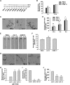
RIC1 Regulates Pollen Tube Germination and Growth. All data are presented as means ± se. *P < 0.05, **P < 0.01, by Student’s t test. (A) COM 1, COM 3, COM 5, COM 6, COM 7, COM 8, COM 11, and COM 12 are transgenic lines generated by transforming ProRIC1:RIC1-GFP into ric1-1 plants. RT-PCR analysis demonstrated that ric1-1 is a null loss-of-function mutant and that RIC1 expression levels in COM 1, COM 5, COM 6, and COM 11 lines were similar to levels in wild-type (Ws-2) plants. EF1αA4 (EF1) served as an internal control. This result is representative of three replicates conducted on three biologically independent samples. (B) The germination frequency of Ws-2, ric1-1, and COM 11 pollen was analyzed at 1 and 1.5 h after the initiation of germination. The germination frequency of COM 11 pollen was similar to that of Ws-2 pollen at each time point. A larger number of ric1-1 pollen grains germinated compared with wild-type pollen at 1 h after initiation. No significant difference was detected at 1.5 h after initiation. About 1000 pollen grains from three independent experiments were analyzed. (C) In vitro-germinated pollen tubes from Ws-2, ric1-1, and COM 11 pollen at 3 h after germination. Bar = 100 μm. (D) The average pollen tube lengths of Ws-2, ric1-1, and COM 11 pollen (only germinated pollen grains were taken into account) were analyzed at 1.5, 3, and 5 h after germination. Pollen tubes from ric1-1 pollen were consistently longer than those of wild-type pollen. The average pollen tube length of COM 11 was similar to Ws-2 at each time point (∼300 pollen tubes per data set). (E) Representative Ws-2, ric1-1, and COM 11 pollen tubes were tracked for 5 min at 2 h after the initiation of germination. The ric1-1 pollen tube grew faster than the Ws-2 and COM 11 tubes. Bar = 10 μm. (F) Pollen tube growth rates were quantitatively analyzed by tracking individual pollen tubes at 2 to 4 h after the initiation of germination for 5 min. The average growth rate of ric1-1 tubes was higher than that of Ws-2 tubes. However, there was no significant difference between Ws-2 and COM 11 tubes (n > 70 pollen tubes per data set). (G) Pollen tubes from wild-type (Col-0) and RIC1 OX (OX 9 and OX 15) pollen were observed at 5 h after germination. Overexpression of RIC1 dramatically inhibited pollen germination. Arrows indicate germinated pollen tubes. Bar = 100 μm. (H) Quantitative real-time PCR analysis showed that the expression of RIC1 is ∼2.5-fold higher in RIC1 OX 9 and OX 15 plants compared with wild-type plants. (I) and (J) The average germination frequency (I) and pollen tube length (J) of Col-0 and RIC1 OX pollen were analyzed 5 h after germination. Overexpression of RIC1 significantly reduced pollen germination as well as pollen tube elongation. Approximately 1000 pollen grains were analyzed to calculate the pollen germination frequency of wild-type and RIC1 OX pollens (I), and ∼200 Col-0 and 30 RIC1 OX pollen tubes were measured for pollen tube length experiments (J).
Figure 2.
Figure 2.
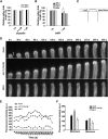
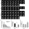
RIC1 Acts on Apical Actin Filaments in Pollen Tubes. All data are presented as means ± se. *P < 0.05, by Student’s t test. (A) Ws-2 and ric1-1 pollen tubes were incubated on germination medium containing different concentrations of oryzalin. The average lengths of treated pollen tubes relative to untreated controls at 3 h after germination were calculated. Untreated controls were normalized to 100% for Ws-2 and ric1-1 pollen. Oryzalin had little effect on pollen tube growth. CK, untreated control. (B) Ws-2, ric1-1, Col-0, and OX 9 pollen tubes were incubated on germination medium containing 1 nM LatB. The average lengths of treated pollen tubes relative to untreated controls at 3 h after germination were calculated. Untreated controls were normalized to 100% for pollen from each line. ric1-1 pollen tubes were more resistant to LatB than wild-type (Ws-2) tubes, whereas there was no significant difference between OX 9 and Col-0 pollen tubes. (C) Schematic showing how the apical dome zone in the pollen tube was defined. A semielliptical region circumscribed by the dotted line at the tip was selected for the apical F-actin analyses shown in (D). (D) Time-lapse images from Supplemental Movie 1 (Col-0), Supplemental Movie 2 [ric1-1 (Col-0)], and Supplemental Movie 3 (RIC1 OX 9) showing F-actin dynamics in growing pollen tubes. Bar = 5 μm. (E) Oscillations of fine F-actin in the apical dome were quantified by measuring fluorescence intensity in representative growing Col-0, ric1-1 (Col-0), and RIC1 OX 9 pollen tubes shown in (D). Compared with Col-0, apical F-actin structures in ric1-1 pollen tubes were more abundant, while there were fewer apical F-actin structures in RIC1 OX 9 pollen tubes. a.u., arbitrary units. (F) Average maximum and minimum fluorescence intensities of F-actin in the apical dome of Col-0 (n = 20), ric1-1 (Col-0) (n = 20), and RIC1 OX 9 (n = 10) pollen tubes. Compared with Col-0, apical F-actin structures in ric1-1 pollen tubes were more abundant with greater oscillatory amplitude, while there were fewer apical F-actin structures with decreased oscillatory amplitude in RIC1 OX 9 pollen tubes.
Figure 3.
Figure 3.
Subcellular Localization of RIC1 Changes during Pollen Tube Germination and Elongation. Pollen grains from COM 11 plants (expressing ProRIC1:RIC1-GFP) were germinated in vitro and observed using spinning disc confocal microscopy. Before germination, RIC1-GFP was detected in the cytoplasm. RIC1-GFP subsequently localized to the PM at the germination site and was restricted to the PM of the growing pollen tube tip. Images were captured every 1 min for 81 min. Bar = 10 μm.
Figure 4.
Figure 4.
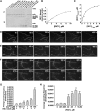
RIC1 Binds and Severs F-Actin in the Presence of Ca2+ in Vitro. All data are presented as means ± se. **P < 0.01, by Student’s t test. (A) A cosedimentation assay was performed to investigate whether RIC1 binds directly to F-actin. Various concentrations of recombinant RIC1 cosedimented with F-actin. Preformed F-actin was incubated with various concentrations of recombinant 6xHis-RIC1. VLN2 served as a positive control. RIC1 in the absence of F-actin and His-GFP served as negative controls. P, pellet; S, supernatant. (B) Densitometry analysis of the results shown in (A). Binding to F-actin was saturated at a stoichiometry of 0.75 mol RIC1/mol G-actin. (C) The amount of RIC1 in the pellet (bound) was plotted versus the amount of RIC1 in the supernatant (free) and fitted with a hyperbolic function. The calculated Kd value was 0.76 μM for this representative experiment. The Kd from three experimental replicates was 0.74 ± 0.08 μM. (D) and (E) Time-lapse images show that RIC1 has no effect on F-actin filament length in the absence of Ca2+. F-actin (polymerized from 0.1 μM rhodamine-labeled G-actin) was incubated with either 100 μM Ca2+ (D) or 0.1 μM RIC1 (E). No obvious changes in F-actin length were observed. Bar = 5 μm for (D) and (E). (F) Time-lapse images from Supplemental Movie 4. Preformed rhodamine-labeled F-actin was monitored by TIRFM immediately after the addition of 0.05 μM RIC1 and 100 μM Ca2+. Individual F-actin filaments exhibited a notable increase in the number of breaks as time elapsed. Bar = 5 μm. (G) F-actin severing frequencies (breaks·μm−1·s−1) in the presence of 0.05 μM RIC1 and various concentrations of Ca2+ were measured (n = ∼25 actin filaments per data set). (H) Mean severing frequencies in the presence of various concentrations of RIC1 and 100 μM Ca2+ were plotted. Actin incubated in the presence of 0.002 μM VLN2 served as a positive control (n = ∼25 actin filaments per experiment).
Figure 5.
Figure 5.
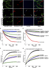
RIC1 Caps the Barbed Ends of F-Actin in Vitro. (A) Dual-color actin filaments displaying polarity. Prepolymerized actin filaments were labeled with Alexa-488 phalloidin and used as seeds. Rhodamine-labeled actin was added to generate dual-color actin filaments with red barbed ends. Bar = 5 μm for (A) to (C). (B) His-RIC1 (arrows) associated with the barbed ends of dual-color actin filaments. His-RIC1 was visualized by staining with anti-His antibody and a Dylight 405-conjugated secondary antibody. (C) Similar associations were not observed in samples that were stained with secondary antibody alone. (D) F-actin was diluted with G buffer in the presence of various concentrations of RIC1. RIC1 prevented actin depolymerization from the barbed ends in a dose-dependent manner. A single representative experiment (n = 3) is shown. a.u., arbitrary units. (E) Preformed F-actin was diluted with G buffer containing 0.12 μM RIC1 in the presence of various concentrations of free Ca2+. RIC1-induced reduction of F-actin depolymerization was significantly enhanced with increasing Ca2+ concentrations. The red trace shows the negative control in which F-actin was diluted with G buffer alone. (F) and (G) Preformed F-actin seeds were incubated with various concentrations of RIC1 (F) or with 0.8 μM RIC1 in the presence of various concentrations of free Ca2+ (G), and 1 μM 10% pyrene-labeled G-actin saturated with 3 μM human profilin I was added to initiate actin elongation at the barbed end. Polymerization was monitored by tracking the increase in pyrene-actin fluorescence upon assembly. A reaction without RIC1 and free Ca2+ served as a negative control (black traces). RIC1 reduced the initial rate of elongation in a dose-dependent manner, and increasing Ca2+ concentrations significantly enhanced the RIC1-induced reduction of F-actin elongation.
Figure 6.
Figure 6.
Quantitative Analysis of Apical/Subapical F-Actin in Pollen Tubes. All data are presented as means ± se. *P < 0.05, **P < 0.01, by Student’s t test. (A) F-actin dynamics were visualized by expressing Lifeact-mEGFP in Col-0, OX 9, and ric1-1 (Col-0) pollen tubes. Images were taken at 2-s intervals. Asterisks indicate individual F-actin filaments initiated from the PM. Arrows indicate F-actin severing events. Bars = 2 μm. (B) The lifetimes of individual PM-associated F-actin filaments were analyzed in Col-0, ric1-1 (Col-0), and OX 9 pollen tubes. Approximately 80 actin filaments from 20 pollen tubes were monitored for each line. The distributions of the lifetimes are presented as the percentage of F-actin lifetimes falling within five ranges (0 to 2, 2 to 6, 6 to 10, 10 to 14, and >14 s). Filament lifetimes were longer in ric1-1 (Col-0) pollen tubes and shorter in RIC1 OX 9 pollen tubes than in wild-type tubes. (C) The average maximum length of single PM-associated F-actin filaments was analyzed in Col-0, ric1-1 (Col-0), and OX 9 pollen tubes. Approximately 80 actin filaments from 20 pollen tubes were monitored for each line. The maximum length reached by single F-actin filaments was longer in ric1-1 (Col-0) pollen tubes and shorter in RIC1 OX 9 tubes compared with wild-type tubes. (D) The average severing frequency of actin filaments was analyzed in Col-0, ric1-1 (Col-0), and OX 9 pollen tubes. The break of actin filaments from the PM was considered the severing event. Approximately 10 pollen tubes were monitored for each line.
Figure 7.
Figure 7.
The Distribution of RIC1 at the PM Oscillates during Pollen Tube Growth. A quantitative analysis of the localization of RIC1-GFP to the apical PM in growing pollen tubes is shown. (A) Schematic illustrating how the amount of apical PM-localized RIC1-GFP was measured. A thin black line was superimposed on the PM. RIC1-GFP localization is represented by thick gray shading. The fluorescence intensity associated with the black line was measured to assess the amount of apical PM-localized RIC1-GFP. (B) Oscillations in the growth rate and the intensity of PM-localized RIC1-GFP were measured in the growing tube shown in Supplemental Movie 8. a.u., arbitrary units. (C) Oscillations in the growth rate and the intensity of PM-localized RIC1-GFP were measured in a tube that was treated with −20°C for 6 min and subsequently warmed to room temperature to resume growth (shown in Supplemental Movie 9). Recovery of RIC1-GFP oscillation precedes the recovery of growth rate oscillation.
Figure 8.
Figure 8.
PM Localization of RIC1 Is Important for Its Function in Regulating Pollen Tube Growth. All data are presented as means ± se. *P < 0.05, **P < 0.01, by Student’s t test. (A) RIC1H37D/H40D contains two point mutations in conserved residues (His-37 and His-40) within the CRIB motif. GFP-tagged RIC1H37D/H40D localized exclusively to the cytoplasm in pollen tubes. Bar = 5 μm. (B) ProLat52:RIC1 or ProLat52:RIC1H37D/H40D was transiently expressed with ProLat52:GFP in tobacco pollen tubes. Pollen tubes transiently expressing ProLat52:GFP served as controls. The average lengths of pollen tubes were measured 5 h after bombardment. Expression of RIC1 resulted in a dramatic reduction of pollen tube growth. Although the expression of RIC1H37D/H40D also reduced pollen tube growth, the degree of reduction was significantly less than that caused by RIC1-GFP. n > 90 pollen tubes per transformation. CK, control pollen tubes expressing ProLat52:GFP only. (C) Localization of stably expressed RIC1-GFP and RIC1H37D/H40D-GFP expressed under the control of the RIC1 native promoter or the Lat52 promoter in Arabidopsis pollen tubes. RIC1-GFP localized to the apical PM under the control of both promoters. RIC1H37D/H40D-GFP was present only in the cytoplasm. Arabidopsis pollen tubes stably expressing ProLat52:GFP served as a control. Bar = 5 μm. (D) Constructs containing GFP, RIC1-GFP, and RIC1H37D/H40D-GFP under the control of the Lat52 promoter were stably transformed into ric1-1 plants. Overexpression of RIC1-GFP dramatically reduced pollen germination as well as pollen tube growth (pollen tubes indicated by arrows). Overexpression of RIC1H37D/H40D-GFP minimally affected pollen germination. Bar = 100 μm. (E) Quantification of the fluorescence intensity associated with pollen grains from ProLat52:GFP (ric1-1), ProLat52:RIC1-GFP (ric1-1), and ProLat52:RIC1H37D/H40D-GFP (ric1-1) plants as shown in (D). Although the fluorescence intensity in ProLat52:RIC1-GFP (ric1-1) pollen grains was much lower (indicating a lower relative expression level) than that of ProLat52:GFP (ric1-1) and ProLat52:RIC1H37D/H40D-GFP (ric1-1) pollen, expression of RIC1-GFP resulted in greater inhibitory effects than expression of RIC1H37D/H40D-GFP (D). a.u., arbitrary units. (F) Average lengths of pollen tubes from the transgenic lines shown in (D). Overexpression of RIC1H37D/H40D-GFP reduced pollen tube growth, but less severely than RIC1-GFP overexpression. n = ∼300 pollen tubes per line. (G) to (I) F-actin stained with Alexa-488 phalloidin was incubated alone (G), with 0.2 μM RIC1H37D/H40D (H), or with 0.2 μM RIC1H37D/H40D and 0.5 μM free Ca2+ (I). The H37D/H40D point mutation did not affect the F-actin bundling and severing activity of RIC1. Bar in (G) = 5 μm for (G) to (I).
Figure 9.
Figure 9.
Different Impacts of RIC1H37D/H40D and RIC1 Overexpression on the Organization and Dynamics of Apical F-Actin. All data are presented as means ± se. *P < 0.05, **P < 0.01, by Student’s t test. (A) Time-lapse images were taken from tobacco pollen tubes 2 to 4 h after particle bombardment, showing F-actin dynamics in growing tobacco pollen tubes. CK, control pollen tubes transiently expressing ProLat52:Lifeact-mEGFP only. Bar = 5 μm. (B) Oscillations in apical fine F-actin were quantified by measuring fluorescence intensity in growing tobacco pollen tubes, shown in (A). a.u., arbitrary units. (C) Average maximum and minimum fluorescence intensities of F-actin in the apical region of tobacco pollen tubes expressing ProLat52:Lifeact-mEGFP only (n = 13), ProLat52:Lifeact-mEGFP with ProLat52:RIC1 (n = 11), or ProLat52:Lifeact-mEGFP with ProLat52:RIC1H37D/H40D (n = 7). The decrease in fluorescence intensity caused by RIC1H37D/H40D overexpression was less severe than the reduction in intensity due to overexpressing wild-type RIC1.
Figure 10.
Figure 10.
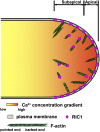
Working Model of RIC1-Mediated Regulation of F-Actin Dynamics in the Tips of Pollen Tubes. Actin filament formation is initiated at the PM in the tip region of the pollen tube. PM-localized RIC1 severs F-actin from the PM, resulting in its dissociation, and facilitates rapid F-actin turnover in the pollen tube apex. The PM distribution of RIC1 oscillates during pollen tube growth, indicating that RIC1 moves between the PM and the cytoplasm. Cytoplasmic RIC1 is still capable of severing F-actin and, therefore, contributes to the formation and dynamics of apical/subapical F-actin structures in pollen tubes. RIC1 also caps the barbed ends of actin filaments to prevent elongation, further contributing to the regulation of the abundance of F-actin in the tip of the pollen tube.

Similar articles

Cited by

References

    1. Boavida L.C., McCormick S. (2007). Temperature as a determinant factor for increased and reproducible in vitro pollen germination in Arabidopsis thaliana. Plant J. 52: 570–582. - PubMed
    1. Bou Daher F., Geitmann A. (2011). Actin is involved in pollen tube tropism through redefining the spatial targeting of secretory vesicles. Traffic 12: 1537–1551. - PubMed
    1. Cai G., Faleri C., Del Casino C., Emons A.M., Cresti M. (2011). Distribution of callose synthase, cellulose synthase, and sucrose synthase in tobacco pollen tube is controlled in dissimilar ways by actin filaments and microtubules. Plant Physiol. 155: 1169–1190. - PMC - PubMed
    1. Cheung A.Y., Wu H.M. (2004). Overexpression of an Arabidopsis formin stimulates supernumerary actin cable formation from pollen tube cell membrane. Plant Cell 16: 257–269. - PMC - PubMed
    1. Cheung A.Y., Wu H.M. (2008). Structural and signaling networks for the polar cell growth machinery in pollen tubes. Annu. Rev. Plant Biol. 59: 547–572. - PubMed

Publication types

MeSH terms

Substances

LinkOut - more resources