. 2015 Jun;8(6):518-27.
doi: 10.1158/1940-6207.CAPR-14-0121. Epub 2015 Mar 24.

Effects of metformin, buformin, and phenformin on the post-initiation stage of chemically induced mammary carcinogenesis in the rat

Affiliations

Effects of metformin, buformin, and phenformin on the post-initiation stage of chemically induced mammary carcinogenesis in the rat

Zongjian Zhu et al. Cancer Prev Res (Phila). 2015 Jun.

Abstract

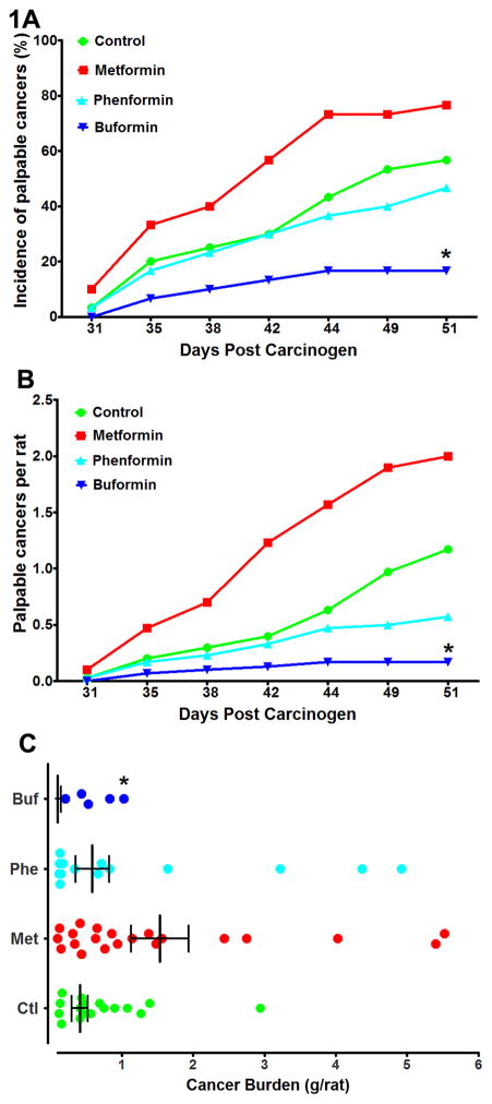
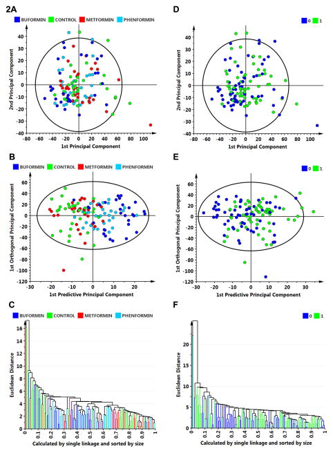
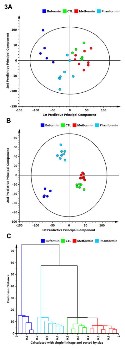
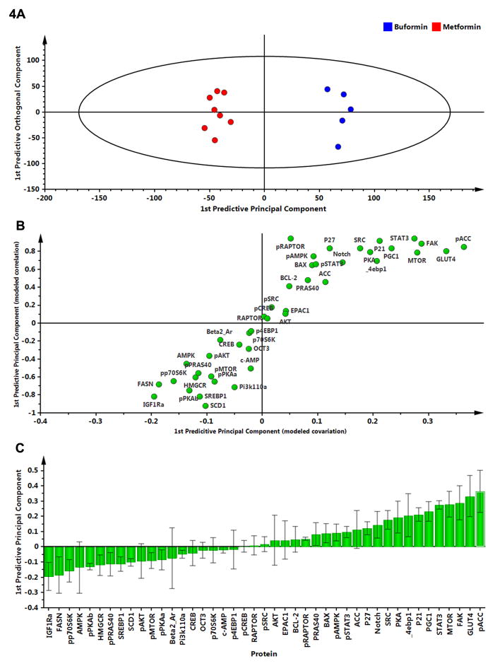
Metformin is a widely prescribed drug for the treatment of type II diabetes. Although epidemiologic data have provided a strong rationale for investigating the potential of this biguanide for use in cancer prevention and control, uncertainty exists whether metformin should be expected to have an impact in nondiabetic patients. Furthermore, little attention has been given to the possibility that other biguanides may have anticancer activity. In this study, the effects of clinically relevant doses of metformin (9.3 mmol/kg diet), buformin (7.6 mmol/kg diet), and phenformin (5.0 mmol/kg diet) were compared with rats fed control diet (AIN93-G) during the post-initiation stage of 1-methyl-1-nitrosourea-induced (50 mg/kg body weight) mammary carcinogenesis (n = 30/group). Plasma, liver, skeletal muscle, visceral fat, mammary gland, and mammary carcinoma concentrations of the biguanides were determined. In comparison with the control group, buformin decreased cancer incidence, multiplicity, and burden, whereas metformin and phenformin had no statistically significant effect on the carcinogenic process relative to the control group. Buformin did not alter fasting plasma glucose or insulin. Within mammary carcinomas, evidence was obtained that buformin treatment perturbed signaling pathways related to energy sensing. However, further investigation is needed to determine the relative contributions of host systemic and cell autonomous mechanisms to the anticancer activity of biguanides such as buformin.


Conflict of interest statement

Conflicts of Interest: The authors disclose no potential conflicts of interest.

Figures

Figure 1
Figure 1
Effects of biguanide treatment on various aspects of the carcinogenic response. A, the incidence of palpable mammary cancer as a function of days post carcinogen injection. B, the average number of palpable cancers per rat as a function of days post carcinogen injection. C, the cancer burden in grams per rat determined at necropsy. The graph is a scatter plot showing the median and interquartile range. The computation includes tumor free rats (not shown in the graph). Note that only 5 buformin treated rats had palpable carcinomas, a fact that limited the number of carcinomas that could be Western blotted in this group. *Significantly different from the control group when adjusted for multiple comparisons.
Figure 2
Figure 2
Multivariate discriminant analysis was used to determine whether plasma analyte data could distinguish among treat groups (A–C) or whether an animal was cancer bearing versus cancer free (D–F). A, to visualize inherent clustering patterns, the scatter plot represents unsupervised analysis through the PCA 4-class model. Poor separation of treatment groups is observed. Model fit: R2X(cum)= 0.437, and Q2(cum)= 0.093. B, to determine contributing sources of variation, the scatter plot represents supervised analysis of the 4-class OPLS-DA model, which rotates the model plane to maximize separation due to class assignment. Separation is still poor with an overall misclassification rate of 45%. Model fit: R2Y(cum)= 0.199, Q2Y(cum)= 0.132. C, to visualize the misclassification rate, the dendrogram depicts hierarchical clustering patterns among treatment groups using single linkage and size. D, to visualize inherent clustering patterns, the scatter plot represents unsupervised analysis through the PCA 2-class model. Poor separation of treatment groups is observed for the categories: cancer free = 0 versus cancer bearing = 1. E, to determine contributing sources of variation, the scatter plot represents supervised analysis of the 2-class OPLS-DA model, which rotates the model plane to maximize separation due to class assignment. Separation is still poor with an overall misclassification rate of 55%. Model fit: R2Y(cum) = 0.124, Q2Y(cum) = 0.080. F, to visualize the misclassification rate, the dendrogram depicts hierarchical clustering patterns among treatment groups using single linkage and size.
Figure 3
Figure 3
Multivariate discriminant analysis was used to determine whether Western blot data for 26 proteins assessed in mammary carcinomas (Supplementary Fig. S1 and 2) could distinguish among treat groups. A, to visualize inherent clustering patterns, the scatter plot represents unsupervised analysis through the PCA 4-class model. Separation of treatment groups is observed. Model fit: R2X(cum) = 0.747, with 5 components, and Q2(cum) = 0.155. B, to determine contributing sources of variation, the scatter plot represents supervised analysis of the 4-class OPLS-DA model, which rotates the model plane to maximize separation due to class assignment. Complete separation of the 4 classes was observed. Model fit: R2Y(cum) = 0.984, Q2Y(cum) = 0.963. C, to visualize the misclassification rate, the dendrogram depicts hierarchical clustering patterns among the treatment groups using single linkage and size. Two main clusters comprise 1) buformin versus 2) phenformin, control, metformin.
Figure 4
Figure 4
To determine the proteins responsible for class separation, multivariate analysis was extended to identify influential proteins responsible for the separation between classes. A, a supervised OPLS-DA model was created to compare buformin to metformin; complete separation was observed. B, an S-plot was constructed by plotting modeled correlation in the first predictive principal component against the modeled covariance in the first predictive component. Upper right and lower left regions of the S-plots contain candidate proteins with both high reliability and high magnitude. C, to determine the statistical reliability of the proteins shown in 4B, jack-knifed confidence intervals (JKCI) were created on the magnitude of covariance in the first component for the 26 proteins and sorted in ascending order based on expression in the buformin group; proteins with JKCIs including 0 were not considered to account for separation.

Similar articles

Cited by

References

    1. Azvolinsky A. Repurposing to fight cancer: the metformin-prostate cancer connection. J Natl Cancer Inst. 2014;106:dju030. - PubMed
    1. Yue W, Yang CS, Dipaola RS, Tan XL. Repurposing of metformin and aspirin by targeting AMPK-mTOR and inflammation for pancreatic cancer prevention and treatment. Cancer Prev Res (Phila) 2014 - PubMed
    1. Quinn BJ, Kitagawa H, Memmott RM, Gills JJ, Dennis PA. Repositioning metformin for cancer prevention and treatment. Trends Endocrinol Metab. 2013;24:469–80. - PubMed
    1. Alimova IN, Liu B, Fan Z, Edgerton SM, Dillon T, Lind SE, et al. Metformin inhibits breast cancer cell growth, colony formation and induces cell cycle arrest in vitro. Cell Cycle. 2009;8:909–15. - PubMed
    1. Liu B, Fan Z, Edgerton SM, Deng XS, Alimova IN, Lind SE, et al. Metformin induces unique biological and molecular responses in triple negative breast cancer cells. Cell Cycle. 2009;8:2031–40. - PubMed

Publication types

MeSH terms