. 2015 Apr 1;142(7):1375-86.
doi: 10.1242/dev.117093.

Activin A directs striatal projection neuron differentiation of human pluripotent stem cells

Affiliations

Activin A directs striatal projection neuron differentiation of human pluripotent stem cells

Charles Arber et al. Development. .

Abstract

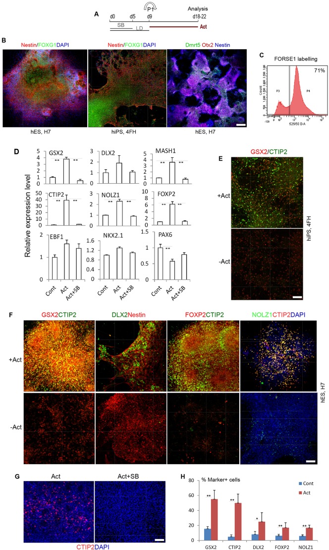
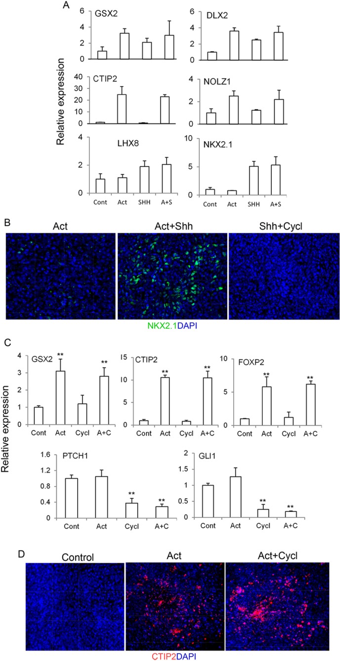
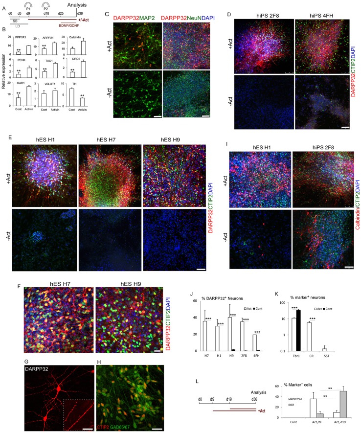
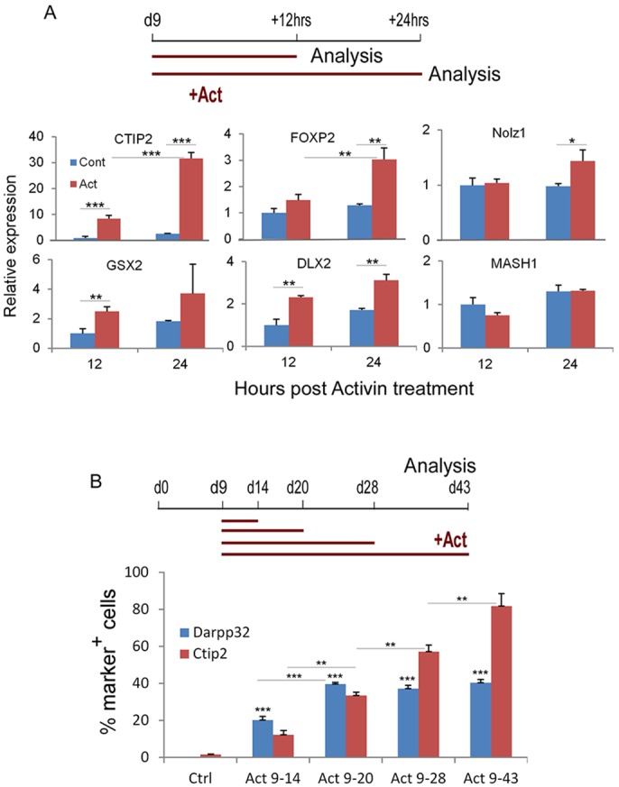
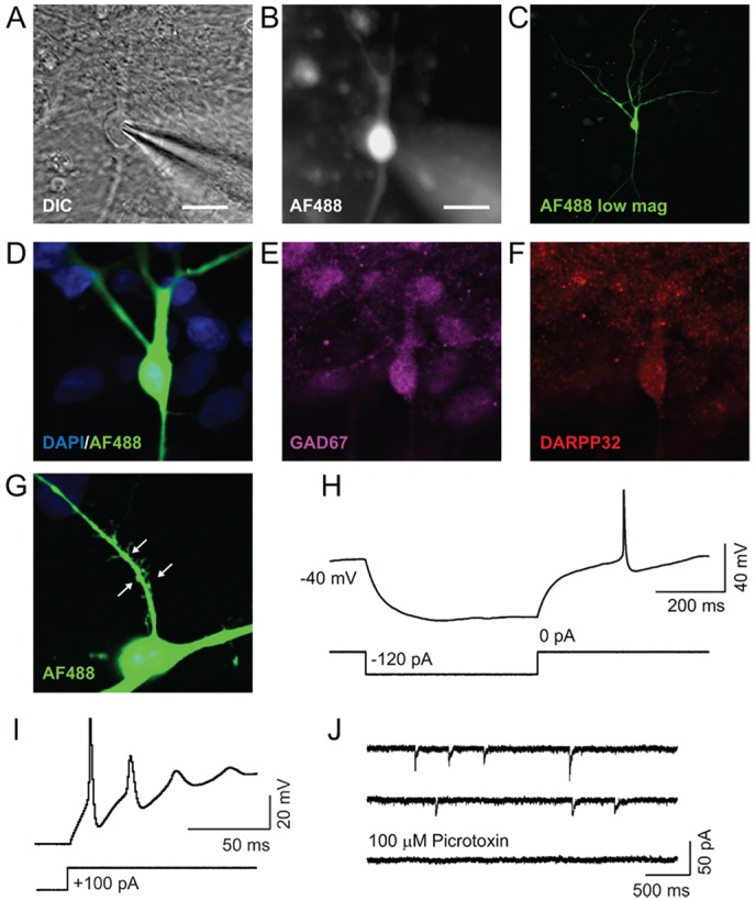
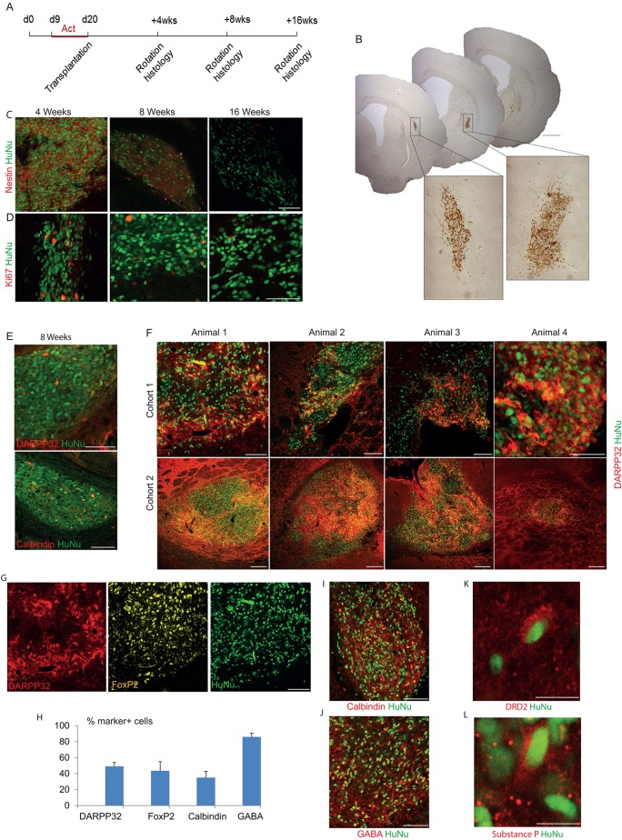
The efficient generation of striatal neurons from human embryonic stem cells (hESCs) and induced pluripotent stem cells (hiPSCs) is fundamental for realising their promise in disease modelling, pharmaceutical drug screening and cell therapy for Huntington's disease. GABAergic medium-sized spiny neurons (MSNs) are the principal projection neurons of the striatum and specifically degenerate in the early phase of Huntington's disease. Here we report that activin A induces lateral ganglionic eminence (LGE) characteristics in nascent neural progenitors derived from hESCs and hiPSCs in a sonic hedgehog-independent manner. Correct specification of striatal phenotype was further demonstrated by the induction of the striatal transcription factors CTIP2, GSX2 and FOXP2. Crucially, these human LGE progenitors readily differentiate into postmitotic neurons expressing the striatal projection neuron signature marker DARPP32, both in culture and following transplantation in the adult striatum in a rat model of Huntington's disease. Activin-induced neurons also exhibit appropriate striatal-like electrophysiology in vitro. Together, our findings demonstrate a novel route for efficient differentiation of GABAergic striatal MSNs from human pluripotent stem cells.

Keywords: Activin; DARPP32 (PPP1R1B); Huntington's disease; Lateral ganglionic eminence; Medium spiny neuron; Neural differentiation; Pluripotent stem cell; Striatum; Transplantation.


Figures

Fig. 1.
Fig. 1.
Activin induces LGE progenitor characteristics and striatal differentiation of hPSCs. (A) Experimental scheme. hESC or hiPSC monolayer cultures were exposed to activin from day 9 to day 18 or 22, when the cultures were harvested for analysis. SB, SB431542; LD, LDN and dorsomorphin; Act, activin A. (B) Antibody staining of a day 18 control culture for the pan-neuroepithelial marker nestin and telencephalic-specific markers FOXG1, OTX2 and cortical neuroepithelial marker DMRT5. Nuclei are counterstained with DAPI (blue). (C) Flow cytometry histogram showing that 71% of the total cell population were FORSE1+ forebrain neural progenitors. (D) qPCR analysis of LGE/striatal and other forebrain regional markers of day 18 cultures following activin treatment from day 9 with or without the Smad inhibitor SB431543, compared with no-activin controls (Cont; set as 1). Data shown are the average of two independent sets of experiments each with duplicate cultures. **P≤0.01, one-way ANOVA followed by Duncan's post-hoc test. (E) Day 18 cultures of 4FH hiPSCs immunostained for GSX2 and CTIP2. (F) Day 22 cultures of H7 hESCs exposed to activin from day 9 and immunostained as indicated. (G) CTIP2 staining of day 20 cultures of H7 cells treated with activin from day 9 in the presence or absence of SB431543 from day 10. (H) Quantification of immunostaining of H7 cells in F. *P≤0.05, **P≤0.01, Student's t-test between control and activin-treated samples for each marker. Scale bars: 100 µm in B; 75 µm in E,F; 60 µm in G.
Fig. 2.
Fig. 2.
Activin induction of LGE characteristics does not require Shh signalling. (A) Day 9 cultures were treated with activin, Shh or both. Cultures were harvested 4 days later for qPCR. (B) NKX2.1 staining of day 20 cultures treated with activin alone (left), activin plus 100 ng/ml Shh, or 100 ng/ml Shh plus 2 µM cyclopamine (Cycl) from day 9. (C) Day 9 cultures were treated with activin, cyclopamine or both. Cultures were harvested 4 days later for qPCR. **P≤0.01, one-way ANOVA. (A,C) Untreated control (Cont) was set to 1. (D) CTIP2 staining of day 20 control culture and cultures treated with activin in the presence or absence of cyclopamine from day 9.
Fig. 3.
Fig. 3.
Efficient generation of striatal projection neurons by activin stimulation. (A) Experimental scheme. (B) qPCR analysis for striatal projection neuron-expressed genes and neuronal transmitter genes. *P≤0.05, **P≤0.01, Student's t-test. (C-I) Immunostaining of day 36-40 cultures for the indicated markers. (G) DARPP32 with a high-magnification inset showing dendrites. (J) Quantification of immunostaining in D,E, showing the percentage of DARPP32+ neurons. (K) Quantification of other neuronal subtypes. ***P≤0.001, Student's t-test between control and activin-treated groups for hPSC lines in J and TBR1 and CR in K. (L) Minimal requirement for activin as evaluated by DARPP32+ neuron production. **P≤0.01, two-way ANOVA. Scale bars: 25 µm in G; 40 µm in C left, F,H; 75 µm in C right, D,E,I.
Fig. 4.
Fig. 4.
Rapid induction and maintenance of CTIP2 by activin. (A) qPCR analysis of LGE/striatal marker genes of day 9 cultures exposed to 12 or 24 h activin treatment, as compared with no-activin controls (12 h value set as 1). Data shown are the average of triplicate cultures from one set of experiments. (B) H9-derived forebrain progenitors were treated with activin from day 9 to day 14, 20, 28 or 43. Cells were fixed at day 43 and immunostained for DARPP32 and CTIP2. P≤0.05, **P≤0.01, ***P≤0.001, two-way ANOVA followed by Bonferroni correction for multiple comparisons.
Fig. 5.
Fig. 5.
Electrophysiological properties of activin-induced GABAergic neurons. (A,B) Differential interference contrast (DIC) image from a whole-cell recording of a differentiated neuron that has been filled with AF488 hydrazide (B). Scale bars: 20 µm. (C) Confocal image of a neuron filled with AF88 hydrazide during recording, and fixed post-recording. (D-F) Confocal images confirming that the recorded neurons (green, D) express the GABAergic markers GAD67 (E) and DARPP32 (F). (G) Magnified view of AF488-filled neuronal spines. Arrows indicate the spiny morphology. (H) Delayed action potential generation was observed after repolarisation from a hyperpolarised state. (I) Evoked action potentials generated in response to a depolarising 100 pA current injection. (J) mIPSCs were observed in the presence of 1 µM tetrodotoxin in the external solution and high [Cl] in the recording pipette. mIPSCs were blocked with 100 µM picrotoxin, confirming their GABAergic identity.
Fig. 6.
Fig. 6.
Robust survival and striatal neuronal differentiation of activin-induced neural progenitors in the adult rat brain. (A) Experimental scheme. (B) Representative low-magnification images of brain sections analysed by HuNu immunohistochemistry at 8 weeks post-transplantation, revealing relatively small grafts with no evidence of overgrowth. Bottom panels are higher magnification photomicrographs of the grafts. (C,D) Microtome sections of brains at 4, 8 and 16 weeks post-transplantation were analysed by immunohistochemistry for HuNu and nestin or Ki67. (E) Immunostaining of 8-week-old grafts for HuNu and DARPP32 or calbindin. (F) DARPP32 and HuNu double staining of 16-week-old grafts. (G) Triple staining for DARPP32, FOXP2 and HuNu on a section of a 16-week-old graft. (H) Quantification of immunostaining of striatal markers, shown as a percentage of HuNu+ cells. (I-L) Immunostaining of 16-week-old grafts for the indicated markers. Unless specified otherwise, images are from 16-week-old grafts. Scale bars: 1 mm in B (low magnification); 100 µm in C-G,I-J; 10 µm in K,L.

Similar articles

Cited by

References

    1. Abdipranoto-Cowley A., Park J. S., Croucher D., Daniel J., Henshall S., Galbraith S., Mervin K. and Vissel B. (2009). Activin A is essential for neurogenesis following neurodegeneration. Stem Cells 27, 1330-1346 10.1002/stem.80 - DOI - PMC - PubMed
    1. Arlotta P., Molyneaux B. J., Jabaudon D., Yoshida Y. and Macklis J. D. (2008). Ctip2 controls the differentiation of medium spiny neurons and the establishment of the cellular architecture of the striatum. J. Neurosci. 28, 622-632 10.1523/JNEUROSCI.2986-07.2008 - DOI - PMC - PubMed
    1. Aubry L., Bugi A., Lefort N., Rousseau F., Peschanski M. and Perrier A. L. (2008). Striatal progenitors derived from human ES cells mature into DARPP32 neurons in vitro and in quinolinic acid-lesioned rats. Proc. Natl. Acad. Sci. USA 105, 16707-16712 10.1073/pnas.0808488105 - DOI - PMC - PubMed
    1. Backman M., Machon O., Mygland L., van den Bout C. J., Zhong W., Taketo M. M. and Krauss S. (2005). Effects of canonical Wnt signaling on dorso-ventral specification of the mouse telencephalon. Dev. Biol. 279, 155-168 10.1016/j.ydbio.2004.12.010 - DOI - PubMed
    1. Cambray S., Arber C., Little G., Dougalis A. G., de Paola V., Ungless M. A., Li M. and Rodríguez T. A. (2012). Activin induces cortical interneuron identity and differentiation in embryonic stem cell-derived telencephalic neural precursors. Nat. Commun. 3, 841 10.1038/ncomms1817 - DOI - PubMed

Publication types

MeSH terms