. 2015 Mar 23;32(6):693-706.
doi: 10.1016/j.devcel.2015.01.028.

A LncRNA-MAF:MAFB transcription factor network regulates epidermal differentiation

Affiliations

A LncRNA-MAF:MAFB transcription factor network regulates epidermal differentiation

Vanessa Lopez-Pajares et al. Dev Cell. .

Abstract

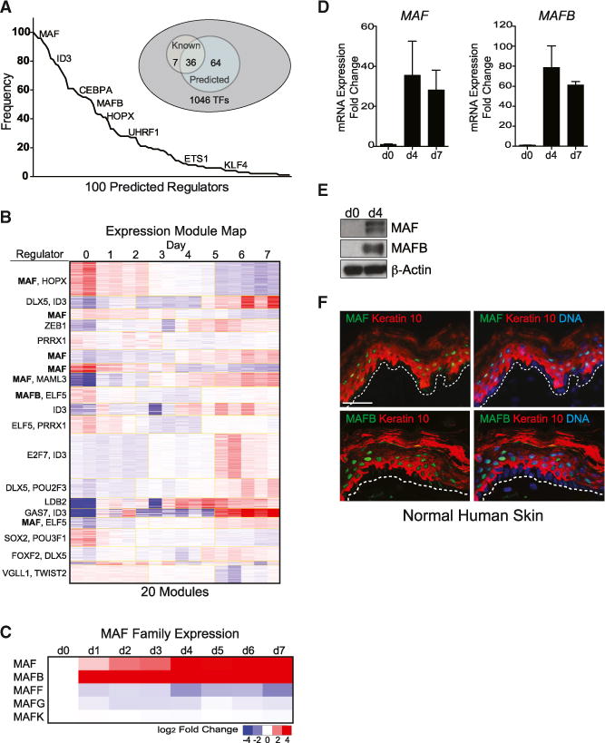
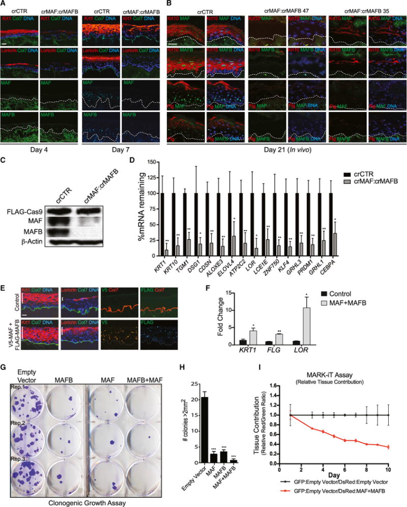
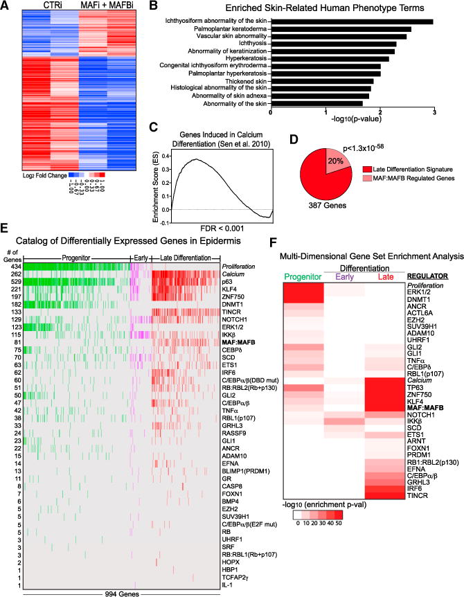
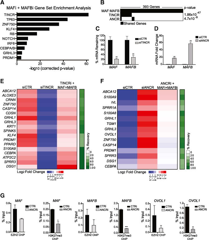
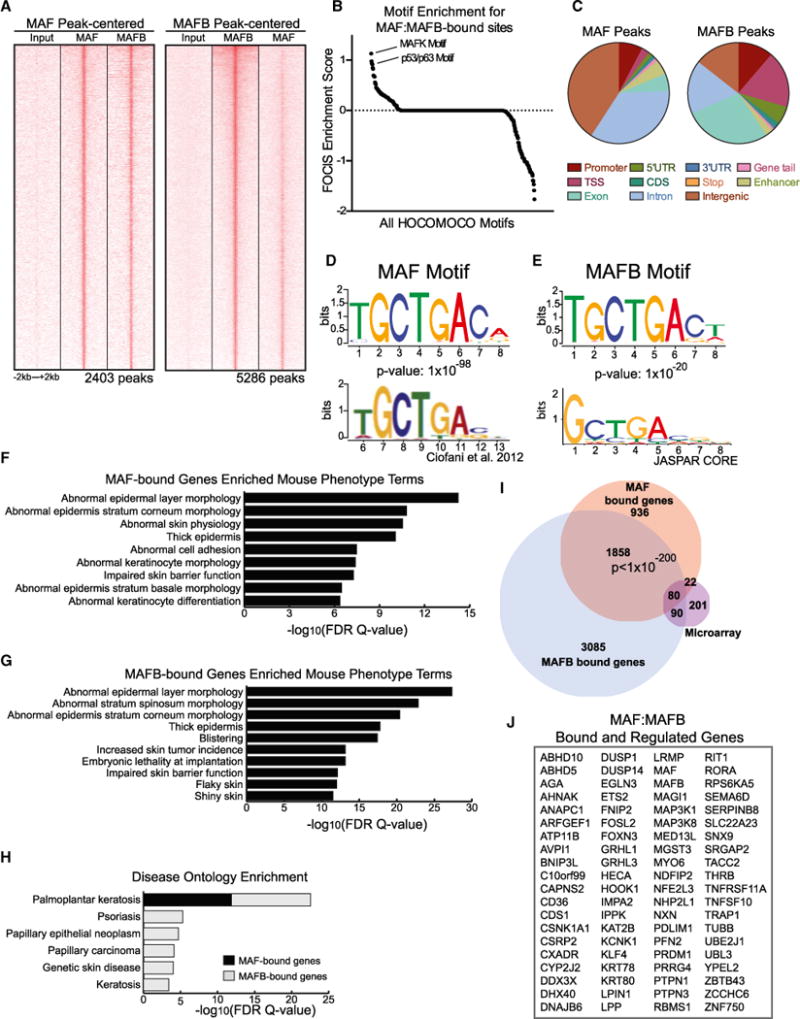
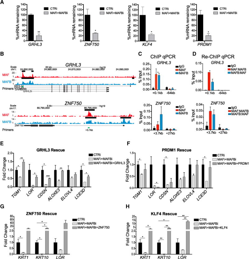
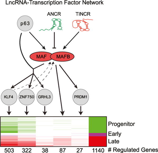
Progenitor differentiation requires remodeling of genomic expression; however, in many tissues, such as epidermis, the spectrum of remodeled genes and the transcription factors (TFs) that control them are not fully defined. We performed kinetic transcriptome analysis during regeneration of differentiated epidermis and identified gene sets enriched in progenitors (594 genes), in early (159 genes), and in late differentiation (387 genes). Module mapping of 1,046 TFs identified MAF and MAFB as necessary and sufficient for progenitor differentiation. MAF:MAFB regulated 393 genes altered in this setting. Integrative analysis identified ANCR and TINCR lncRNAs as essential upstream MAF:MAFB regulators. ChIP-seq analysis demonstrated MAF:MAFB binding to known epidermal differentiation TF genes whose expression they controlled, including GRHL3, ZNF750, KLF4, and PRDM1. Each of these TFs rescued expression of specific MAF:MAFB target gene subsets in the setting of MAF:MAFB loss, indicating they act downstream of MAF:MAFB. A lncRNA-TF network is thus essential for epidermal differentiation.


Figures

Figure 1
Figure 1. MAF and MAFB Are Induced in Epidermal Differentiation
(A) Expression module mapping of time-course microarrays performed in Genomica against a set of 1,046 potential regulators expressed in skin with GO term “transcription factor.” Graph represents frequency of predicted regulators in 100 permutations of module mapping with select TFs displayed. (B) Representative output of expression module map showing high frequency of MAF in predicted module regulation. Yellow boxes demarcate modules. (C–E) (C) Gene expression changes in MAF TF family members during regeneration of differentiated epidermis. Induction of MAF and MAFB (D) mRNA and (E) protein expression during calcium-induced keratinocyte differentiation in vitro. Mean ± SEM; n = 2 biological replicates. (F) MAF and MAFB protein expression (green) in differentiated layers, represented by keratin 10 staining (orange), of normal adult skin tissue (dotted white line demarcates the epidermal basement membrane). Scale bar, 50 mm (see also Figures S1 and S2).
Figure 2
Figure 2. MAF:MAFB Is Necessary and Sufficient for Differentiation Gene Induction and Progenitor Compartment Exit
(A) Organotypic culture of epidermis generated by CRISPR/Cas9-mediated genetic ablation of MAF and MAFB (crMAF:crMAFB) across day 4 and day 7 of differentiation, with staining for differentiation markers (top two rows) keratin 1 and loricrin (orange), collagen VII (green), and nuclei (blue) or staining for (bottom two rows) MAF or MAFB (green). Note loss of differentiation proteins upon MAF:MAFB loss. Scale bar, 50 μm. (B) In vivo skin xenografts generated from CRISPR/Cas9-targeted primary keratinocytes with a control sgRNA or two different combinations of sgRNAs targeting MAF and MAFB (crMAF:crMAFB 47 or crMAF:MAFB 35), harvested 21 days post-seeding, and immunostained for differentiation markers keratin 10 and filaggrin (red) and MAF or MAFB (green). Scale bar, 50 μm. (C) Western blots for Cas9, MAF, and MAFB in CRISPR/Cas9-targeted primary keratinocytes. (D) Quantification of differentiation gene expression levels in crMAF:crMAFB day 7 organotypic tissue. Mean ± SEM; n = 3 biological replicates; *p < 0.05, **p < 0.01. (E) Immunostaining of overexpressed epitope-tagged V5-MAF and FLAG-MAFB in organotypic epidermis and differentiation markers as in (A, top); white brackets indicate increased distribution of differentiation protein expression in MAF:MAFB compared to control (left); epitope tag immunostaining shows MAF and MAFB overexpression throughout the tissue (right). Scale bar, 50 μm. (F) Quantification of mRNA levels of differentiation markers in tissue overexpressing MAF:MAFB. Mean ± SEM; n = 2 biological replicates; *p < 0.05, **p < 0.01. (G) Clonogenic assay of progenitor keratinocytes overexpressing MAF and/or MAFB over a 2-week time span. (H) Quantification of colonies > 2 mm2. Mean ± SEM; n = 4; ***p < 0.001. (I) MARK-iT cell competition assay over 10 days showing relative tissue contribution detected by red:green fluorescence ratios of GFP:empty vector/DsRed:empty vector mixed samples compared to GFP:empty vector/DsRed:MAF+MAFB mixed samples; note progressive decrease in the proportion of MAF:MAFB-expressing cells over time. Mean ± SEM; n = 3 biological replicates (see also Figure S3).
Figure 3
Figure 3. MAF:MAFB Regulates Epidermal Differentiation Genes
(A) Epidermal differentiation genes regulated by MAF:MAFB. Heatmap of duplicate, mean-centered microarray analysis of MAF:MAFB depleted tissue showing genes significantly changed compared to control siRNA. (B) Skin-related human disease phenotype terms of MAF:MAFB target genes. (C) GSEA of MAF:MAFB depleted tissue against a calcium-differentiated keratinocyte gene set (Sen et al., 2010) ranked by MAF:MAFB genes downregulated to upregulated (left to right). (D) Quantification of the overlap of MAF:MAFB downregulated genes with the late differentiation signature. (E) Catalog of published regulators controlling the epidermal progenitor, early and late differentiation gene sets, ranked by the number of genes regulated. (F) Multi-dimensional GSEA conducted on published epidermal regulator gene sets against the three differentiation signatures (-log10 enrichment p value; maximum p < 0.05), with progenitor to late differentiation gene regulation ordered from top to bottom (see also Figure S4).
Figure 4
Figure 4. LncRNAs Are Upstream Regulators of MAF:MAFB
(A) GSEA of genes downregulated in MAF:MAFB-depleted tissue overlaps with gene sets of known epidermal differentiation regulators. (B–D) (B) Overlap of MAFi:MAFBi, TINCRi, and ANCRi gene sets in keratinocytes. Quantification of MAF and MAFB mRNA levels in (C) TINCR-depleted tissue and (D) ANCR-depleted progenitor keratinocytes. Mean ± SEM; n = 2 biological replicates; **p < 0.01. (E) Enforced expression of MAF:MAFB in TINCR-depleted tissue partially rescues the differentiation defect observed with TINCR knockdown; heatmap shows average fold change by qRT-PCR (n = 2; p < 0.05, t test) and percentage recovery of differentiation genes in green bar at right. (F) Concurrent knockdown of MAF and MAFB partially rescues the ANCRi phenotype of premature differentiation gene induction; heatmap shows average fold by qRT-PCR (n = 2; p < 0.05, t test) and percentage recovery of differentiation genes. (G) ChIP-qPCR showing binding of EZH2 at ANCR-regulated genes in CTRi- and ANCRi-treated progenitor keratinocytes and the H3K27me3-repressive histone mark at these loci. Mean ± SEM; n = 2; *p < 0.05, **p < 0.01, ***p < 0.001 (see also Figure S5).
Figure 5
Figure 5. Genome-wide MAF:MAFB Binding in Differentiated Keratinocytes
(A) Heatmap of MAF-centered ChIP-seq peaks aligned with input and MAFB ChIP-seq peaks (left) and MAFB-centered ChIP-seq peaks with input and MAF ChIP-seq peaks (right). (B) FOCIS analysis was used to identify motifs predicted to co-occur within MAF and MAFB peaks; noted are two of the top hits. (C–G) (C) Distribution of MAF peaks (left) and MAFB peaks (right) across the genome. Position-weight matrix of motifs discovered with ChIP-seq peaks for MAF with published (D) MAF motif from Ciofani et al. and MAFB-discovered motif with JASPAR (E) MAFB motif. Enriched mouse phenotype terms for genes associated with (F) MAF ChIP-seq peaks and (G) MAFB ChIP-seq peaks. (H) Disease ontology enrichment observed for genes associated with MAF and MAFB peaks. (I) Venn diagram showing overlap of MAFi:MAFBi microarray genes with MAF-bound and MAFB-bound genes. (J) Eighty genes whose expression changes with knockdown of both MAF and MAFB and that are associated with both MAF and MAFB ChIP-seq peaks (see also Figure S6).
Figure 6
Figure 6. MAF and MAFB Control Differentiation-Inducing Epidermal TFs
(A) mRNA levels of epidermal TFs in MAF:MAFB-depleted epidermal tissue. (B) MAF (top, red) and MAFB (bottom, blue) ChIP-seq tracks at epidermal TF genes, GRHL3 and ZNF750; black bars denote ChIP-seq peaks called by MACS and green bars denote sequences for ChIP-qPCR. (C) ChIP-qPCR of enriched MAF and MAFB peaks at GRHL3 and ZNF750. (D) Re-ChIP-qPCR of enriched MAF and MAFB peaks showing occupancy of both MAF and MAFB on TF gene loci. (E–H) Overexpression of (E) GRHL3, (F) PRDM1, (G) ZNF750, or (H) KLF4 partially rescues impaired differentiation observed in MAFi:MAFBi tissue. Mean ± SEM; n = 2 biological replicates; *p < 0.05, **p < 0.01, ***p < 0.001 (see also Figure S7).
Figure 7
Figure 7. LncRNA:TF Network Regulating Epidermal Differentiation
Proposed model of a lncRNA-MAF:MAFB-TF network regulating epidermal differentiation.

Comment in

References

    1. Artner I, Blanchi B, Raum JC, Guo M, Kaneko T, Cordes S, Sieweke M, Stein R. MafB is required for islet b cell maturation. Proc Natl Acad Sci USA. 2007;104:3853–3858. - PMC - PubMed
    1. Aziz A, Soucie E, Sarrazin S, Sieweke MH. MafB/c-Maf deficiency enables self-renewal of differentiated functional macrophages. Science. 2009;326:867–871. - PubMed
    1. Bao X, Tang J, Lopez-Pajares V, Tao S, Qu K, Crabtree GR, Khavari PA. ACTL6a enforces the epidermal progenitor state by suppressing SWI/SNF-dependent induction of KLF4. Cell Stem Cell. 2013;12:193–203. - PMC - PubMed
    1. Birnbaum RY, Zvulunov A, Hallel-Halevy D, Cagnano E, Finer G, Ofir R, Geiger D, Silberstein E, Feferman Y, Birk OS. Seborrhea-like dermatitis with psoriasiform elements caused by a mutation in ZNF750, encoding a putative C2H2 zinc finger protein. Nat Genet. 2006;38:749–751. - PubMed
    1. Blank V, Andrews NC. The Maf transcription factors: regulators of differentiation. Trends Biochem Sci. 1997;22:437–441. - PubMed

Publication types

MeSH terms

Associated data