. 2015 Mar 24;6(2):e00322-15.
doi: 10.1128/mBio.00322-15.

Type IV pilus secretins have extracellular C termini

Affiliations

Type IV pilus secretins have extracellular C termini

Joshua A Lieberman et al. mBio. .

Abstract

Type IV pili (T4Ps) are surface appendages used by Gram-negative and Gram-positive pathogens for motility and attachment to epithelial surfaces. In Gram-negative bacteria, such as the important pediatric pathogen enteropathogenic Escherichia coli (EPEC), during extension and retraction, the pilus passes through an outer membrane (OM) pore formed by the multimeric secretin complex. The secretin is common to Gram-negative assemblies, including the related type 2 secretion (T2S) system and the type 3 secretion (T3S) system. The N termini of the secretin monomers are periplasmic and in some systems have been shown to mediate substrate specificity. In this study, we mapped the topology of BfpB, the T4P secretin from EPEC, using a combination of biochemical and biophysical techniques that allowed selective identification of periplasmic and extracellular residues. We applied rules based on solved atomic structures of outer membrane proteins (OMPs) to generate our topology model, combining the experimental results with secondary structure prediction algorithms and direct inspection of the primary sequence. Surprisingly, the C terminus of BfpB is extracellular, a result confirmed by flow cytometry for BfpB and a distantly related T4P secretin, PilQ, from Pseudomonas aeruginosa. Keeping with prior evidence, the C termini of two T2S secretins and one T3S secretin were not detected on the extracellular surface. On the basis of our data and structural constraints, we propose that BfpB forms a beta barrel with 16 transmembrane beta strands. We propose that the T4P secretins have a C-terminal segment that passes through the center of each monomer.

Importance: Secretins are multimeric proteins that allow the passage of secreted toxins and surface structures through the outer membranes (OMs) of Gram-negative bacteria. To date, there have been no atomic structures of the C-terminal region of a secretin, although electron microscopy (EM) structures of the complex are available. This work provides a detailed topology prediction of the membrane-spanning domain of a type IV pilus (T4P) secretin. Our study used innovative techniques to provide new and comprehensive information on secretin topology, highlighting similarities and differences among secretin subfamilies. Additionally, the techniques used in this study may prove useful for the study of other OM proteins.


Figures

FIG 1
FIG 1
Iodination of extracellular residues in intact cells. Purified BfpB from labeled intact cells was subjected to MS identification of extracellular residues as indicated by an iodination event. The graph displays the number of iodination events divided by the total number of times the peptide was recovered to yield an iodination percentage.
FIG 2
FIG 2
Solvent accessibility labeling of BfpB-CSM. Western blot results from TMM(PEG)12 labeling of BfpB-CSM. The 50-kDa marker bands are shown in lanes M. (A) Results of TMM(PEG)12 labeling of control BfpB-CSM constructs. After 2-h incubation with the reagent, a second band (solid arrow) with decreased Mr was observed, indicating that a positive label was consistently observed for BfpBT309C and BfpBS41C. No such band was ever observed with either wild-type (WT) BfpB or BfpBC539S. (B) Control to confirm that the slower-migrating band is due to TMM(PEG)12. The Western blot shows the absence of the slower-migrating band without TMM(PEG)12 labeling (open arrow). (C) TMM(PEG)12 labeling of all BfpB-CSM mutants reveals that all but three (wild-type BfpB, BfpBC539S, and BfpBS490C) are labeled and therefore solvent accessible.
FIG 3
FIG 3
Förster resonance energy transfer (FRET) following in vitro labeling of BfpB cysteine mutants. The engineered and single endogenous cysteine residues in BfpB were labeled with TAMRA-maleimide, and the BfpB C terminus was labeled with streptavidin-Alexa Fluor 488. For each construct, FRET efficiency (as a percentage) was calculated from three independent labeling experiments. The primary y axis (left) shows FRET efficiency (black bars), while the secondary y axis (right) shows acceptor fluorescence (in arbitrary units; RFU, relative fluorescence units) (gray bars). Error bars show standard deviations. Asterisks indicate FRET signals significantly different from the wild-type BfpB baseline, as determined by ANOVA.
FIG 4
FIG 4
Flow cytometry of BfpB and PilQ in situ reveals an extracellular C terminus for T4P secretins. Cells expressing either DsbA (negative control), intimin557 (positive control), BfpB, PilQ (P. aeruginosa, T4P), EscC (EPEC, T3S), GspD (enterotoxigenic E. coli, T2S), EpsD (V. cholerae, T2S), or BfpBΔIPS, each with an N-terminal Strep tag, were labeled with SA-488 in triplicate experiments and analyzed by flow cytometry for surface fluorescence. Results are expressed as a percentage of positive cells for each sample.
FIG 5
FIG 5
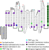
Model topology of BfpB with 16 transmembrane beta strands and an intrapore segment. The predicted topology of BfpB is presented here from the N terminus to the C terminus with the extracellular space at the top of the page and the periplasmic space at the bottom, generated in part using the TOPO2 program (S. J. Johns, TOPO2 transmembrane protein display software [http://www.sacs.ucsf.edu/TOPO2/]). The N-terminal, lipid-anchored cysteine, C18, is colored gold. Tyrosines and histidines labeled exclusively by iodination of purified protein are colored blue, while residues iodinated in intact cells are colored red. Residues that were mutated to cysteines and utilized for FRET experiments, as well as the endogenous C539, are colored by FRET labeling results: light green if FRET negative (neg.) or cyan if FRET positive (pos.). Residues colored black indicate that a cysteine mutation resulted in an unstable BfpB protein, while residues colored gray indicate that a cysteine mutation produced stable BfpB multimers but did not complement the bfpB mutant for autoaggregation. Aromatic residues are colored magenta. The 16th strand contains a putative E. coli Bam-stop signal, consisting of a terminal Phe (98). Beyond the 16th strand is the intrapore segment (IPS), which is predicted to extend from I538 to K548.

References

    1. Craig L, Pique ME, Tainer JA. 2004. Type IV pilus structure and bacterial pathogenicity. Nat Rev Microbiol 2:363–378. doi:10.1038/nrmicro885. - DOI - PubMed
    1. Pelicic V. 2008. Type IV pili: e pluribus unum? Mol Microbiol 68:827–837. doi:10.1111/j.1365-2958.2008.06197.x. - DOI - PubMed
    1. Peabody CR, Chung YJ, Yen MR, Vidal-Ingigliardi D, Pugsley AP, Saier MH. 2003. Type II protein secretion and its relationship to bacterial type IV pili and archaeal flagella. Microbiology 149:3051–3072. doi:10.1099/mic.0.26364-0. - DOI - PubMed
    1. Melville S, Craig L. 2013. Type IV pili in Gram-positive bacteria. Microbiol Mol Biol Rev 77:323–341. doi:10.1128/MMBR.00063-12. - DOI - PMC - PubMed
    1. Fröls S, Ajon M, Wagner M, Teichmann D, Zolghadr B, Folea M, Boekema EJ, Driessen AJ, Schleper C, Albers SV. 2008. UV-inducible cellular aggregation of the hyperthermophilic archaeon Sulfolobus solfataricus is mediated by pili formation. Mol Microbiol 70:938–952. doi:10.1111/j.1365-2958.2008.06459.x. - DOI - PubMed

Publication types

MeSH terms

LinkOut - more resources