

Colonic sarcoidosis: unusual onset of a systemic disease
- PMID: 25805948
- PMCID: PMC4363771
- DOI: 10.3748/wjg.v21.i11.3380
Colonic sarcoidosis: unusual onset of a systemic disease
Abstract
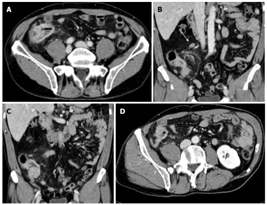
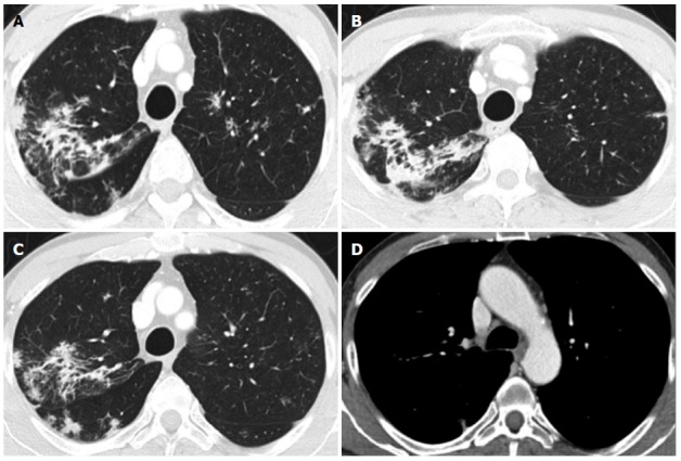
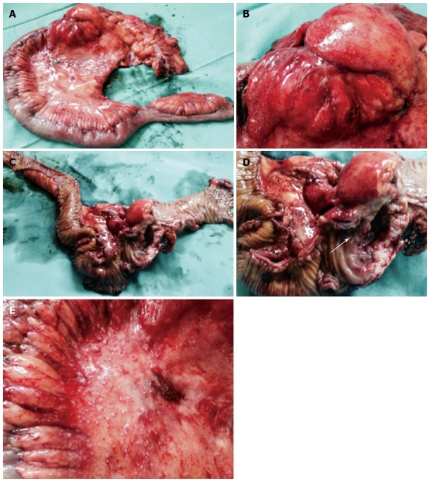
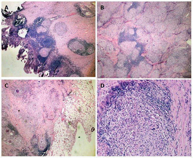
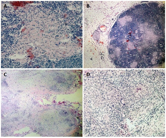
Sarcoidosis is a multisystem chronic inflammatory condition of unknown etiology that has the potential to involve every tissue in the body. Sarcoidosis in the gastrointestinal system, and particularly the colon, is very rare. Here, we report the case of a 57-year-old man with no previous diagnosis of sarcoidosis who presented with new onset of abdominal pain and constipation. A colonoscopy revealed that the abdominal pain was caused by an obstructing lesion in the cecum-ascending colon and lacked a clear histologic diagnosis. Radiologic investigation revealed concentric wall thickening of the cecum-ascending colon with multiple satellite lymphadenopathies, highly suggestive of a malignancy. The patient underwent a laparotomy and a right hemicolectomy was performed. A diagnosis of colonic sarcoidosis was made after the resected specimen was examined. Additionally, a chest computed tomography scan revealed lung involvement with atypical radiologic features in the absence of respiratory symptoms. Only histologic examination of the surgical specimen can yield a diagnosis of gastrointestinal sarcoidosis due to the non-specificity of endoscopic and radiologic findings.
Keywords: Colon; Hemicolectomy; Noncaseating granuloma; Sarcoidosis; Systemic disease.
Figures
References
-
- MacArthur KL, Forouhar F, Wu GY. Intra-abdominal complications of sarcoidosis. J Formos Med Assoc. 2010;109:484–492. - PubMed
-
- Statement on sarcoidosis. Joint Statement of the American Thoracic Society (ATS), the European Respiratory Society (ERS) and the World Association of Sarcoidosis and Other Granulomatous Disorders (WASOG) adopted by the ATS Board of Directors and by the ERS Executive Committee, February 1999. Am J Respir Crit Care Med. 1999;160:736–755. - PubMed
-
- Henke CE, Henke G, Elveback LR, Beard CM, Ballard DJ, Kurland LT. The epidemiology of sarcoidosis in Rochester, Minnesota: a population-based study of incidence and survival. Am J Epidemiol. 1986;123:840–845. - PubMed
-
- Lynch JP, Kazerooni EA, Gay SE. Pulmonary sarcoidosis. Clin Chest Med. 1997;18:755–785. - PubMed
-
- Baughman RP, Teirstein AS, Judson MA, Rossman MD, Yeager H, Bresnitz EA, DePalo L, Hunninghake G, Iannuzzi MC, Johns CJ, et al. Clinical characteristics of patients in a case control study of sarcoidosis. Am J Respir Crit Care Med. 2001;164:1885–1889. - PubMed
Publication types
MeSH terms
LinkOut - more resources
Full Text Sources
Other Literature Sources
Medical
