Review
. 2015 Mar 9:9:59.
doi: 10.3389/fnins.2015.00059. eCollection 2015.

Features of alpha-synuclein that could explain the progression and irreversibility of Parkinson's disease

Affiliations
Review

Features of alpha-synuclein that could explain the progression and irreversibility of Parkinson's disease

Scarlet Gallegos et al. Front Neurosci. .

Abstract

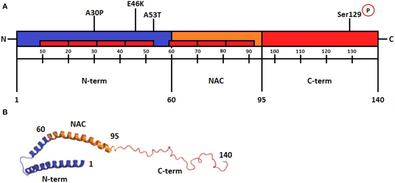
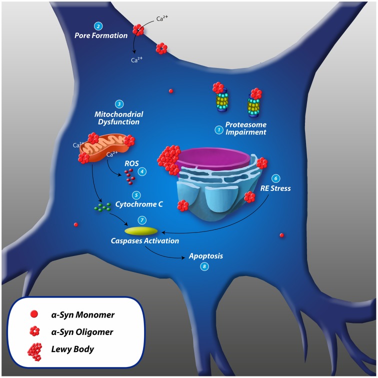
Alpha-synuclein is a presynaptic protein expressed throughout the central nervous system, and it is the main component of Lewy bodies, one of the histopathological features of Parkinson's disease (PD) which is a progressive and irreversible neurodegenerative disorder. The conformational flexibility of α-synuclein allows it to adopt different conformations, i.e., bound to membranes or form aggregates, the oligomers are believed to be the more toxic species. In this review, we will focus on two major features of α-synuclein, transmission and toxicity, that could help to understand the pathological characteristics of PD. One important feature of α-synuclein is its ability to be transmitted from neuron to neuron using mechanisms such as endocytosis, plasma membrane penetration or through exosomes, thus propagating the Lewy body pathology to different brain regions thereby contributing to the progressiveness of PD. The second feature of α-synuclein is that it confers cytotoxicity to recipient cells, principally when it is in an oligomeric state. This form causes mitochondrial dysfunction, endoplasmic reticulum stress, oxidative stress, proteasome impairment, disruption of plasma membrane and pore formation that lead to apoptosis pathway activation and consequent cell death. The complexity of α-synuclein oligomerization and formation of toxic species could be a major factor for the irreversibility of PD and could also explain the lack of successful therapies to halt the disease.

Keywords: Parkinson's disease; alpha-synuclein; neurodegeneration; toxicity; transmission.


Figures

Figure 1
Figure 1
(A) Schematic representation of α-synuclein regions. The amino-terminal from amino acids 1–60 is an amphipathic region responsible for α-syn-membrane interactions. It contains repeats of hexamer motifs similar to apolipoproteins (represented in the figure as red rectangles). The point mutations of α-syn are located in this region (A30P, E46K, and A53T). The central region from amino acids 61-95, termed NAC (non-β amyloid component), is the most hydrophobic portion of the protein and is required for the aggregation process. This region folds into a β-sheet secondary structure and forms amyloid fibrils. The C-terminal from amino acids 96–140 is characterized by the presence of acidic residues and several negative charges. The residue serine 129 in this region is phosphorylated in Lewy bodies (Modified from Plotegher et al., 2014). (B) Schematic representation of micelle-bound α-synuclein. The N-terminal region with antiparallel α-helices is shown in blue, the NAC region is also an α-helix and is shown in orange, and the unstructured C-terminal part is shown in red. Numbers refer to amino acid residues. (Modified from Lashuel et al., 2013). Reprinted by permission from MacMillan Publishers Ltd (Nature Reviews Neuroscience).
Figure 2
Figure 2
Cell-to-cell transmission mechanisms of α-synuclein. (1) Exosomes are small membrane vesicles derived from the endocytic pathway, and the presence of α-syn inside exosomes and its transmission to recipient cells has been reported. (2) α-syn could be released through an exocytosis process and accumulate in the extracellular space and be taken up by other cells through endocytosis. (3) α-syn associates to biological membranes by its N-terminal, penetrates the membrane and gains access to the cytosol. (4) Tunneling nanotubes are membrane bridges of actin between cells, but this mechanism has not been identified to participate in α-syn transmission. (5) Dying cells could be an important reservoir of pathological α-syn when they eventually expel their contents after being lysed.
Figure 3
Figure 3
Toxic mechanisms of α-synuclein and cell death. (1) Proteasome impairment: inhibition of chymotryptic, tryptic and post-acidic proteasome activity leading to further intracellular accumulation of misfolded proteins such as α-syn. (2) Pore formation: α-syn oligomers penetrate cellular membranes increasing the conductance by forming pore-like structures that could act as non-selective channels, resulting in abnormal calcium influx. (3) Mitochondrial dysfunction: α-syn associates to the mitochondrial inner and outer membrane and (4) Increases mitochondrial and intracellular ROS levels. (5) Release of cytochrome C: accumulation of intramitochondrial ROS and Ca2+ leads to reduction in mitochondrial membrane potential and opening of permeability transition pores that could cause the release of cytochrome C to the cytosol. (6) RE stress: cellular accumulation of misfolded proteins can lead to chronic endoplasmic reticulum stress. α-syn associates to ER membrane and causes morphologic dysfunction such as dilated cisternae and increases the level of ER chaperones. (7) Cytochrome C leads to activation of caspase-3 and -9, and ER stress leads to activation of caspase-12. (8) Caspases initiate apoptosis leading to cell death.

Similar articles

Cited by

References

    1. Aguayo L. G., Parodi J., Sepúlveda F. J., Opazo C. (2009). Pore-forming neurotoxin-like mechanism for Aβ oligomer-induced synaptic failure, in Pore-Forming Neurotoxin-Like Mechanism for Aβ Oligomer-Induced Synaptic Failure, eds Perry G., Maccioni R. B. (New York, NY: Springer; ), 13–21 10.1007/978-0-387-87995-6 - DOI
    1. Ahn K. J., Paik S. R., Chung K. C., Kim J. (2006). Amino acid sequence motifs and mechanistic features of the membrane translocation of alpha-synuclein. J. Neurochem. 97:265279. 10.1111/j.1471-4159.2006.03731.x - DOI - PubMed
    1. Alvarez-Erviti L., Seow Y., Schapira A. H., Gardiner C., Sargent I. L., Wood M. J., et al. . (2011). Lysosomal dysfunction increases exosome-mediated alpha-synuclein release and transmission. Neurobiol. Dis 42, 360–367. 10.1016/j.nbd.2011.01.029 - DOI - PMC - PubMed
    1. Angot E., Brundin P. (2009). Dissecting the potential molecular mechanisms underlying α-synuclein cell-to-cell transfer in Parkinson's disease. Parkinsonism Relat. Disord. 15, S143–S147. 10.1016/S1353-8020(09)70802-8 - DOI - PubMed
    1. Angot E., Steiner J. A., Lema Tome C. M., Ekstrom P., Mattsson B., Bjorklund A., et al. . (2012). Alpha-synuclein cell-to-cell transfer and seeding in grafted dopaminergic neurons in vivo. PloS ONE 7:e39465. 10.1371/journal.pone.0039465 - DOI - PMC - PubMed

LinkOut - more resources