

Cognitive training-induced short-term functional and long-term structural plastic change is related to gains in global cognition in healthy older adults: a pilot study
- PMID: 25805989
- PMCID: PMC4353252
- DOI: 10.3389/fnagi.2015.00014
Cognitive training-induced short-term functional and long-term structural plastic change is related to gains in global cognition in healthy older adults: a pilot study
Abstract
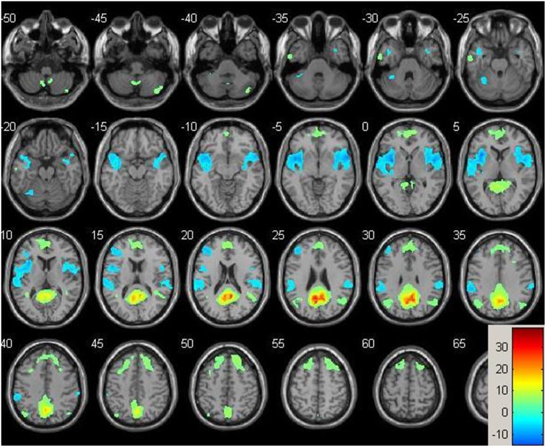
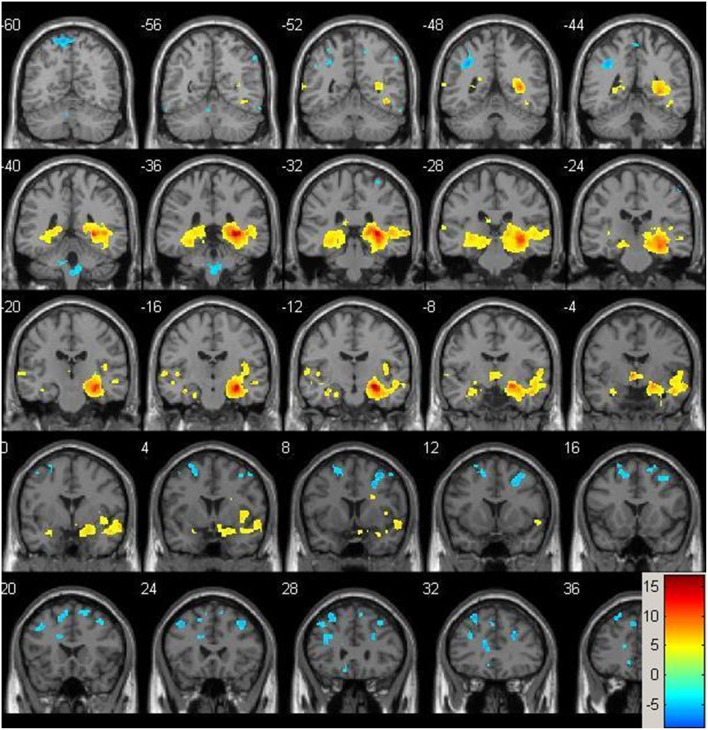
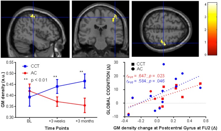
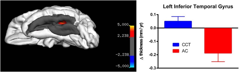
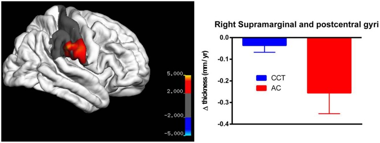
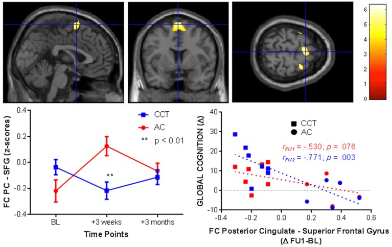
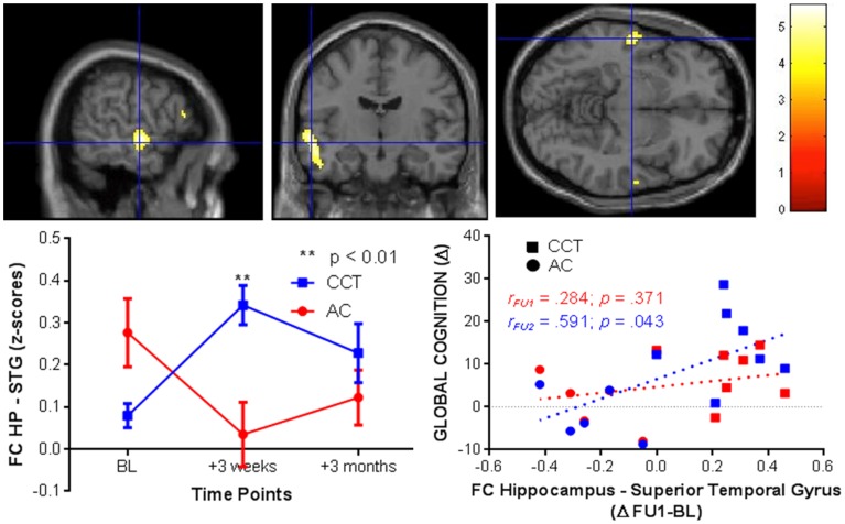
Computerized cognitive training (CCT) is a safe and inexpensive intervention to enhance cognitive performance in the elderly. However, the neural underpinning of CCT-induced effects and the timecourse by which such neural changes occur are unknown. Here, we report on results from a pilot study of healthy older adults who underwent three 1-h weekly sessions of either multidomain CCT program (n = 7) or an active control intervention (n = 5) over 12 weeks. Multimodal magnetic resonance imaging (MRI) scans and cognitive assessments were performed at baseline and after 9 and 36 h of training. Voxel-based structural analysis revealed a significant Group × Time interaction in the right post-central gyrus indicating increased gray matter density in the CCT group compared to active control at both follow-ups. Across the entire sample, there were significant positive correlations between changes in the post-central gyrus and change in global cognition after 36 h of training. A post-hoc vertex-based analysis found a significant between-group difference in rate of thickness change between baseline and post-training in the left fusiform gyrus, as well as a large cluster in the right parietal lobe covering the supramarginal and post-central gyri. Resting-state functional connectivity between the posterior cingulate and the superior frontal gyrus, and between the right hippocampus and the superior temporal gyrus significantly differed between the two groups after 9 h of training and correlated with cognitive change post-training. No significant interactions were found for any of the spectroscopy and diffusion tensor imaging data. Though preliminary, our results suggest that functional change may precede structural and cognitive change, and that about one-half of the structural change occurs within the first 9 h of training. Future studies are required to determine the role of these brain changes in the mechanisms underlying CCT-induced cognitive effects.
Keywords: cognitive training; healthy older adults; hippocampus; magnetic resonance imaging; post-central gyrus; posterior cingulate.
Figures

Similar articles
-
Analysis of central mechanism of cognitive training on cognitive impairment after stroke: Resting-state functional magnetic resonance imaging study.J Int Med Res. 2014 Jun;42(3):659-68. doi: 10.1177/0300060513505809. Epub 2014 Apr 10. J Int Med Res. 2014. PMID: 24722262 Clinical Trial.
-
Therapeutically relevant structural and functional mechanisms triggered by physical and cognitive exercise.Mol Psychiatry. 2016 Nov;21(11):1633-1642. doi: 10.1038/mp.2016.19. Epub 2016 Mar 22. Mol Psychiatry. 2016. PMID: 27001615 Free PMC article.
-
[Diffusion tensor imaging and resting-state functional magnetic resonance imaging in patients with delirium in intensive care unit].Zhonghua Wei Zhong Bing Ji Jiu Yi Xue. 2020 Jan;32(1):88-93. doi: 10.3760/cma.j.cn121430-20190905-00016. Zhonghua Wei Zhong Bing Ji Jiu Yi Xue. 2020. PMID: 32148238 Chinese.
-
Functional brain changes associated with cognitive training in healthy older adults: A preliminary ALE meta-analysis.Brain Imaging Behav. 2020 Aug;14(4):1247-1262. doi: 10.1007/s11682-019-00080-0. Brain Imaging Behav. 2020. PMID: 30900077 Review.
-
Immediate and long-term efficacy of executive functions cognitive training in older adults: A systematic review and meta-analysis.Psychol Bull. 2019 Jul;145(7):698-733. doi: 10.1037/bul0000196. Epub 2019 Apr 18. Psychol Bull. 2019. PMID: 30998045
Cited by
-
MIND food and speed of processing training in older adults with low education, the MINDSpeed Alzheimer's disease prevention pilot trial.Contemp Clin Trials. 2019 Sep;84:105814. doi: 10.1016/j.cct.2019.105814. Epub 2019 Jul 18. Contemp Clin Trials. 2019. PMID: 31326523 Free PMC article.
-
A Large-Scale, Cross-Sectional Investigation Into the Efficacy of Brain Training.Front Hum Neurosci. 2019 Jul 9;13:221. doi: 10.3389/fnhum.2019.00221. eCollection 2019. Front Hum Neurosci. 2019. PMID: 31338032 Free PMC article.
-
Microscopic Fractional Anisotropy Detects Cognitive Training-Induced Microstructural Brain Changes.Tomography. 2022 Jan 1;8(1):33-44. doi: 10.3390/tomography8010004. Tomography. 2022. PMID: 35076639 Free PMC article. Clinical Trial.
-
Early-Life Cognitive Activity Is Related to Reduced Neurodegeneration in Alzheimer Signature Regions in Late Life.Front Aging Neurosci. 2018 Mar 22;10:70. doi: 10.3389/fnagi.2018.00070. eCollection 2018. Front Aging Neurosci. 2018. PMID: 29623037 Free PMC article.
-
Does Functional Connectivity Provide a Marker for Cognitive Rehabilitation Effects in Alzheimer's Disease? An Interventional Study.J Alzheimers Dis. 2017;57(4):1303-1313. doi: 10.3233/JAD-160773. J Alzheimers Dis. 2017. PMID: 28372326 Free PMC article. Clinical Trial.
References
-
- Barnes D. E., Santos-Modesitt W., Poelke G., Kramer A. F., Castro C., Middleton L. E., et al. . (2013). The Mental Activity and eXercise (MAX) Trial: a randomized controlled trial to enhance cognitive function in older adults. JAMA Intern. Med. 173, 797–804. 10.1001/jamainternmed.2013.189 - DOI - PMC - PubMed
LinkOut - more resources
Full Text Sources
Other Literature Sources
Medical