. 2012 Feb 5;7(4):295-301.
doi: 10.3969/j.issn.1673-5374.2012.04.010.

Erythropoietin upregulates growth associated protein-43 expression and promotes retinal ganglion cell axonal regeneration in vivo after optic nerve crush

Affiliations

Erythropoietin upregulates growth associated protein-43 expression and promotes retinal ganglion cell axonal regeneration in vivo after optic nerve crush

Haibo Tan et al. Neural Regen Res. .

Abstract

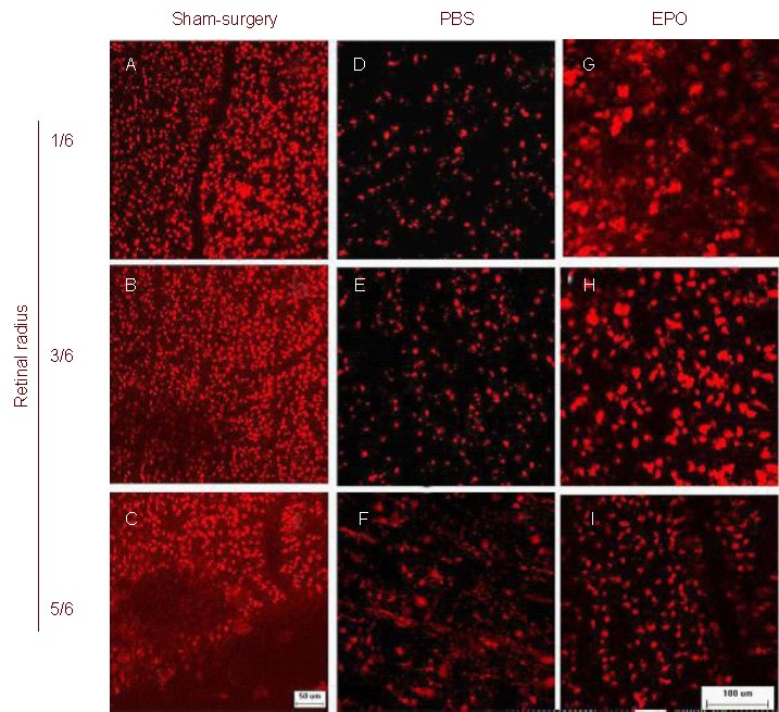
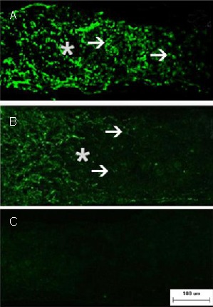
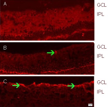
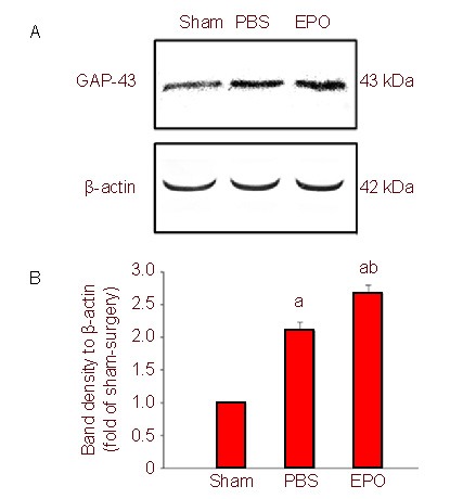
In this study, we established a rat model of optic nerve crush to explore the effects of erythropoietin on retinal ganglion cell axonal regeneration. At 15 days after injury in erythropoietin treated rats, retinal ganglion cell densities in regions corresponding to the 1/6, 3/6 and 5/6 ratios of the retinal radius were significantly increased. In addition, the number of growth associated protein-43 positive axons was significantly increased at different distances (50, 250 and 500 μm) from the crush site after erythropoietin treatment. Erythropoietin significantly increased growth associated protein-43 protein levels in the retina after crush injury, as determined by western blot and immunofluorescence analysis. These results demonstrate that erythropoietin protects injured retinal ganglion cells and promotes axonal regeneration.

Keywords: axonal regeneration; erythropoietin; neural regeneration; optic nerve crush; retinal ganglion cells.


Conflict of interest statement

Conflicts of interest: None declared.

Figures

Figure 1
Figure 1
Morphology of RGCs after optic nerve crush (DiI staining; scale bar: 50 μm in A–C and 100 μm in D–I. Representative photographs of the flat-mounted retinas at the corresponding areas (at 1/6, 3/6 and 5/6 of the retinal radius) showed DiI-labeled RGCs in retinas on day 15 after injury. Almost all RGCs were labeled and the morphology of the RGCs was round in the sham-surgery (A–C) and EPO (G–I) groups. In the PBS group (D–F), there were a few DiI-labeled RGCs and microglia were obviously labeled at 1/6 and 3/6 radius, and were more numerous at 5/6 retinal radius. RGCs: Retinal ganglion cells; Dil: 1, 1-dioctadecyl-3, 3, 3′, 3′-tetramethyl-indocarbocyanine perchlorate; PBS: phosphate buffered saline; EPO: erythropoietin.
Figure 2
Figure 2
Axonal regeneration in retinal ganglion cells after optic nerve crush (immunofluorescence staining; fluorescein isothiocyanate-labeling; scale bar: 100 μm). Representative photomicrographs show the crush site (asterisk) and regenerating axons with growth associated protein-43 immunofluorescence (arrowheads) on day 15 after injury. Numerous growth associated protein-43 positive axons are seen passing through the crush site and they grew beyond the distal crush site in the EPO group (A). In contrast, very few axons traversed the crush site in the PBS group (B). In addition, the sham-surgery group (C) showed no staining. PBS: Phosphate buffered saline; EPO: erythropoietin.
Figure 3
Figure 3
GAP-43 expression in the retina after optic nerve crush (immunofluorescence; Cy3-labeled; scale bars: 10 µm in (A–B) and 20 μm in (C). (A) Sham-surgery group; (B) PBS group; (C) EPO group. The retina in the sham-surgery group showed little GAP-43 expression. In contrast, GAP-43 expression was intense (arrowhead, red fluorescence) in the PBS group. The immunoreactivity of GAP-43 (arrowheads, red fluorescence) in the EPO group was greater than in the PBS group. GAP-43: Growth associated protein-43; GCL: ganglion cell layer; IPL: inner plexiform layer; PBS: phosphate buffered saline; EPO: erythropoietin.
Figure 4
Figure 4
Growth associated protein-43 (GAP-43) expression in retina after optic nerve crush (western blot). (A) Representative western blots for GAP-43 in the retina. Erythropoietin (EPO) treatment resulted in significantly increased GAP-43 expression. (B) Quantification of western blot bands for GAP-43 protein levels from six independent experiments. aP < 0.01, vs. sham-surgery (sham) group; bP < 0.01, vs. phosphate buffered saline (PBS) group (one-way analysis of variance).

References

    1. Tang Z, Zhang S, Lee C, et al. An optic nerve crush injury murine model to study retinal ganglion cell survival. J Vis Exp. 2011;50:2685. - PMC - PubMed
    1. Yoles E, Schwartz M. Degeneration of spared axons following partial white matter lesion: implications for optic nerve neuropathies. Exp Neurol. 1998;153(1):1–7. - PubMed
    1. Levin LA. Axonal loss and neuroprotection in optic neuropathies. Can J Ophthalmol. 2007;42(3):403–408. - PubMed
    1. Zalish M, Lavie V, Duvdevani R, et al. Gangliosides attenuate axonal loss after optic nerve injury. Retina. 1993;13(2):145–147. - PubMed
    1. Fitzgerald M, Payne SC, Bartlett CA, et al. Secondary retinal ganglion cell death and the neuroprotective effects of the calcium channel blocker lomerizine. Invest Ophthalmol Vis Sci. 2009;50(11):5456–5462. - PubMed