Review
. 2015 Jan 30:5:5.
doi: 10.4103/2156-7514.150448. eCollection 2015.

Transcatheter renal interventions: a review of established and emerging procedures

Affiliations
Review

Transcatheter renal interventions: a review of established and emerging procedures

Jeet Minocha et al. J Clin Imaging Sci. .

Abstract

Catheter-based interventions play an important role in the multidisciplinary management of renal pathology. The array of procedures available to interventional radiologists (IRs) includes established techniques such as angioplasty, stenting, embolization, thrombolysis, and thrombectomy for treatment of renovascular disease, as well as embolization of renal neoplasms and emerging therapies such as transcatheter renal artery sympathectomy for treatment of resistant hypertension. Here, we present an overview of these minimally invasive therapies, with an emphasis on interventional technique and clinical outcomes of the procedure.

Keywords: Angioplasty; catheter; embolization; renal; stenting.


Conflict of interest statement

Conflict of Interest: None declared.

Figures

Figure 1
Figure 1
78-year-old woman with medically refractory hypertension and renal artery stenosis. (a) Abdominal aortogram exhibits stenosis of proximal renal arteries bilaterally (arrowheads). (b) Right renal arteriogram better delineates stenosis (arrowheads). (c) Final right renal arteriogram performed after primary stent deployment shows marked improvement in the renal artery caliber (arrowheads).
Figure 2
Figure 2
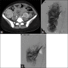
40-year-old man suffered penetrating flank trauma resulting in renal artery pseudoaneurysm. (a) Contrast-enhanced axial CT image reveals left peri-renal hematoma (arrowheads) and focal round high attenuation pseudoaneurysm (arrow). (b) Main left renal arteriogram and (c) selective left renal arteriogram demonstrate traumatic renal artery pseudoaneurysm (arrowheads). Note the presence of arteriovenous fistula, with early filling of renal vein (arrows). (d) Completion arteriogram following coil embolization (arrowhead) of feeding artery shows no filling of pseudoaneurysm.
Figure 3
Figure 3
60-year-old woman with intermittent right flank pain found to have congenital renal arteriovenous malformation. (a) Right renal arteriogram demonstrates arteriovenous malformation (arrowheads). Note hypertrophy of main renal artery (black arrow) and early renal venous drainage (white arrow). (b) Final right renal arteriogram following vascular plug (arrowhead) embolization displays no residual filling of the renal arteriovenous malformation.
Figure 4
Figure 4
32-year-old asymptomatic woman with incidental discovery of renal angiomyolipoma. (a) Contrast-enhanced axial CT image in tuberous sclerosis patient demonstrates 5.8 cm right lower pole renal AML (arrow). (b) Right renal arteriogram displays multiple renal masses (arrowheads), consistent with the patient's known AMLs, including lower pole mass (white arrowheads). (c) Post-embolization right renal arteriogram shows no significant residual flow to embolized renal AMLs; in this case, particles and metallic coils were used.
Figure 5
Figure 5
48-year-old man with flank pain found to have renal cell carcinoma. (a) Contrast-enhanced axial CT image demonstrates 19 cm left renal mass (arrowheads); preoperative embolization requested prior to resection. (b) Left renal arteriogram displays hypervascular tumor (arrowheads) with abundant neovascularity. (c) Post-embolization left renal arteriogram following particle embolization shows no significant residual flow to tumor, with pruning of distal vasculature; metallic coils placed in distal renal artery after particle devascularization (not shown).

References

    1. Martin LG, Rundback JH, Wallace MJ, Cardella JF, Angle JF, Kundu S, et al. Society of Interventional Radiology (SIR). Quality improvement guidelines for angiography, angioplasty, and stent placement for the diagnosis and treatment of renal artery stenosis in adults. J Vasc Interv Radiol. 2010;21:421–30. - PubMed
    1. Prince EA, Murphy TP, Hampson CO. Catheter-based arterial sympathectomy: Hypertension and beyond. J Vasc Interv Radiol. 2012;23:1125–34. - PubMed
    1. Conlon PJ, Athirakul K, Kovalik E, Schwab SJ, Crowley J, Stack R, et al. Survival in renal vascular disease. J Am Soc Nephrol. 1998;9:252–6. - PubMed
    1. Isles CG, Robertson S, Hill D. Management of renovascular disease: A review of renal artery stenting in ten studies. QJM. 1999;92:159–67. - PubMed
    1. Rees CR. Stents for atherosclerotic renovascular disease. J Vasc Interv Radiol. 1999;10:689–705. - PubMed

LinkOut - more resources