Case Reports
. 2015 Jan 30:5:8.
doi: 10.4103/2156-7514.150454. eCollection 2015.

Hypertrophic olivary degeneration - a report of two cases

Affiliations
Case Reports

Hypertrophic olivary degeneration - a report of two cases

Radha Sarawagi et al. J Clin Imaging Sci. .

Abstract

Hypertrophic olivary degeneration (HOD) is seen following lesions in the Guillain-Mollaret triangle. This is unique because the inferior olivary nucleus hypertrophies following degeneration unlike the typical atrophy seen in other structures. We report two cases of HOD in two different clinical scenarios.

Keywords: Anterolateral medulla; Guillain–Mollaret triangle; T2-hyperintensity; hypertrophic olivary degeneration.


Conflict of interest statement

Conflict of Interest: None declared.

Figures

Figure 1
Figure 1
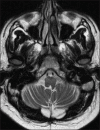
Case 1. 42-year-old male with bilateral hypertrophic olivary degeneration following excision of a fourth ventricular epidermoid cyst Axial. T2-weighted images through fourth ventricle (a and b) show postoperative gliosis (white arrows).
Figure 2
Figure 2
Case 1. 42-year-old male with bilateral hypertrophic olivary degeneration following excision of a fourth ventricular epidermoid cyst Axial. T2-weighted images (a and b) through medulla show bilateral hyperintensities in olivary region with mild hypertrophy (white arrows).
Video 1
Video 1
42-year-old male with bilateral hypertrophic olivary degeneration following excision of a fourth ventricular epidermoid cyst. Video clip shows palatal myoclunus.
Figure 3
Figure 3
Case 1. 42-year-old male with bilateral hypertrophic olivary degeneration following excision of a fourth ventricular epidermoid cyst. Axial T2-weighted image 14 months later through same level as 2(b) shows reduction in hypertrophy and hyperintensity.
Figure 4
Figure 4
Case 2. 49-year-old male with unilateral HOD following brainstem hemorrhage. (a) and (b) T2-weighted axial images through medulla show ill-defined hyperintensities in olivary region on right side with mild hypertrophy (white arrows).
Figure 5
Figure 5
Case 2. 49-year-old male with unilateral hypertrophic olivary degeneration following brainstem hemorrhage. (a) Axial T2-weighted gradient image through midbrain shows susceptibility effect suggestive of chronic hemorrhage on right side involving the region of red nucleus (white arrow). (b) T2-weighted coronal images through medulla shows ill-defined hyperintensity in olivary region on right side (red arrow). Evidence of hemorrhage is noted as heterogeneous hypointensity in right side of pons (white arrow).
Figure 6
Figure 6
(a) Illustrative sketch of the Guillain–Mollaret triangle shows the inferior olivary nucleus (in blue), contralateral dentate nucleus of the cerebellum (in green) and the ipsilateral red nucleus (in red). (b) Schematic diagram of Guillain–Mollaret triangle, superimposed on T2W coronal image.

References

    1. Sanverdi SE, Oguz KK, Haliloglu G. Hypertrophic olivary degeneration in children: Four new cases and a review of the literature with an emphasis on the MRI findings. Br J Radiol. 2012;85:511–6. - PMC - PubMed
    1. Krings T, Foltys H, Meister IG, Reul J. Hypertrophic olivary degeneration following pontine haemorrhage: Hypertensive crisis or cavernous haemangioma bleeding? J Neurol Neurosurg Psychiatry. 2003;74:797–9. - PMC - PubMed
    1. Kitajima M, Korogi Y, Shimomura O, Sakamoto Y, Hirai T, Miyayama H, et al. Hypertrophic olivary degeneration: MR imaging and pathologic findings. Radiology. 1994;192:539–43. - PubMed
    1. Vaidhyanath R, Thomas A, Messios N. Bilateral hypertrophic olivary degeneration following surgical resection of a posterior fossa epidermoid cyst. Br J Radiol. 2010;83:e211–5. - PMC - PubMed
    1. Goyal M, Versnick E, Tuite P, Cyr JS, Kucharczyk W, Montanera W, et al. Hypertrophic olivary degeneration: Metaanalysis of the temporal evolution of MR findings. AJNR Am J Neuroradiol. 2000;21:1073–7. - PMC - PubMed

Publication types