

Biofilms in infections of the eye
- PMID: 25806622
- PMCID: PMC4384075
- DOI: 10.3390/pathogens4010111
Biofilms in infections of the eye
Abstract
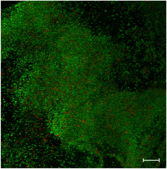
The ability to form biofilms in a variety of environments is a common trait of bacteria, and may represent one of the earliest defenses against predation. Biofilms are multicellular communities usually held together by a polymeric matrix, ranging from capsular material to cell lysate. In a structure that imposes diffusion limits, environmental microgradients arise to which individual bacteria adapt their physiologies, resulting in the gamut of physiological diversity. Additionally, the proximity of cells within the biofilm creates the opportunity for coordinated behaviors through cell-cell communication using diffusible signals, the most well documented being quorum sensing. Biofilms form on abiotic or biotic surfaces, and because of that are associated with a large proportion of human infections. Biofilm formation imposes a limitation on the uses and design of ocular devices, such as intraocular lenses, posterior contact lenses, scleral buckles, conjunctival plugs, lacrimal intubation devices and orbital implants. In the absence of abiotic materials, biofilms have been observed on the capsule, and in the corneal stroma. As the evidence for the involvement of microbial biofilms in many ocular infections has become compelling, developing new strategies to prevent their formation or to eradicate them at the site of infection, has become a priority.
Figures
References
-
- Westall F., Witb M.J., Dannb J., van der Gaastc S., de Ronded C.E.J., Gernekee D. Early archean fossil bacteria and biofilms in hydrothermally-influenced sediments from the barberton greenstone belt, south africa. Precambrian Res. 2001;106:93–116. doi: 10.1016/S0301-9268(00)00127-3. - DOI
Publication types
Grants and funding
LinkOut - more resources
Full Text Sources
Other Literature Sources
Molecular Biology Databases
