Case Reports
. 2015 Mar 26;372(13):1223-30.
doi: 10.1056/NEJMoa1407647.

Human infection with a zoonotic orthopoxvirus in the country of Georgia

Affiliations
Case Reports

Human infection with a zoonotic orthopoxvirus in the country of Georgia

Neil M Vora et al. N Engl J Med. .

Abstract

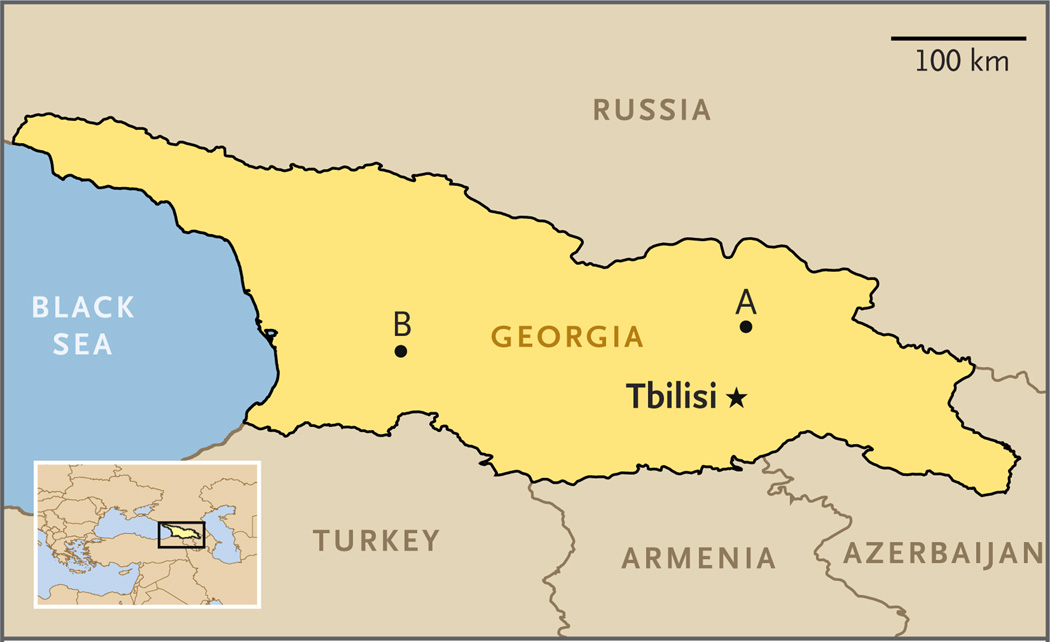
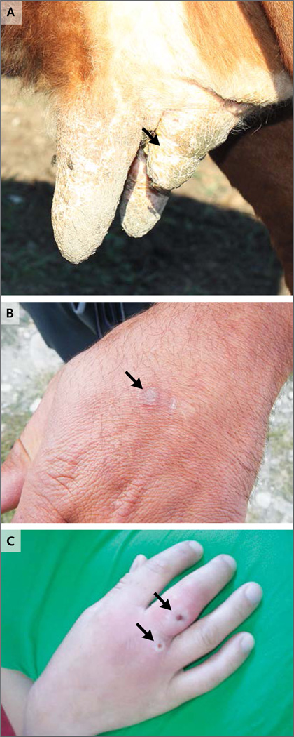
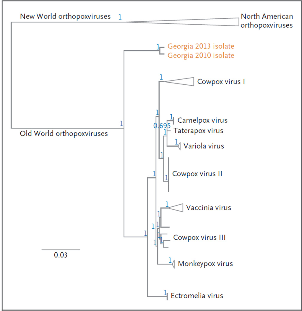
During 2013, cutaneous lesions developed in two men in the country of Georgia after they were exposed to ill cows. The men had never received vaccination against smallpox. Tests of lesion material with the use of a quantitative real-time polymerase-chain-reaction assay for non-variola virus orthopoxviruses were positive, and DNA sequence analysis implicated a novel orthopoxvirus species. During the ensuing epidemiologic investigation, no additional human cases were identified. However, serologic evidence of exposure to an orthopoxvirus was detected in cows in the patients' herd and in captured rodents and shrews. A third case of human infection that occurred in 2010 was diagnosed retrospectively during testing of archived specimens that were originally submitted for tests to detect anthrax. Orthopoxvirus infection should be considered in persons in whom cutaneous lesions develop after contact with animals.


Figures

Figure 1
Figure 1. Location of Human Infections with the Novel Orthopoxvirus in Georgia
The location marked A indicates where the two index patients began to have symptoms in 2013. The location marked B indicates where samples were obtained in 2010 from the patient in whom infection was diagnosed retrospectively.
Figure 2
Figure 2. Clinical Manifestations and Sequelae of Infection with the Novel Orthopoxvirus
Panel A shows a cow from the index herd tended to by the two index patients. The cow had lesions on its teats before the patients’ illnesses. After the cow’s illness, a previously normal teat became contracted (arrow) and had decreased milk production. Anti-orthopoxvirus IgG was detected in a serum sample obtained from this cow at an end-point titer of 1:400 or higher. Panel B shows a healed lesion (arrow) on the left hand of the first index patient approximately 3 months after the onset of illness. Panel C shows active lesions (arrows) on the right hand of a woman who received a retrospective diagnosis of infection with the novel virus. This photograph (courtesy of Maka Tsilosani) was taken in 2010 when the woman was ill.
Figure 3
Figure 3. Phylogenetic Analysis of the Novel Orthopoxvirus
The phylogenetic tree was constructed with the use of sequence data generated from the viruses that infected the patients in 2010 and 2013. The loci that were concatenated for phylogenetic analysis were vaccinia virus Copenhagen homologues A7L, A10L, A24R, D1R, D5R, E6R, E9L, H4L and J6R. The alignment was calculated with the use of Geneious software, version 6.1.2 (Biomatters); the Translation Align and ClustalW alignment options were implemented. The phylogenetic tree was inferred with the use of the MrBayes plug-in software, version 3.2.1, with a general time-reversible substitution model, inverse-gamma rate variation, chain length of 5 million generations, burn-in length of 1 million generations, subsampling every 1000 generations, and unconstrained branch lengths. The positions of the Georgian isolates were established by performing three successive phylogenetic analyses, from the subfamily level (Chordopoxvirinae) to the genus level (orthopoxvirus). Triangles indicate collapsed branches with a common origin. The position of the isolates from Georgia represents a deep divergence relative to other Eurasian isolates, suggesting a remote common ancestry with variola, monkeypox, vaccinia, and cowpox viruses (which probably diverged from one another more recently). The blue numbers at each node correspond to posterior probability values for each group. The scale bar corresponds to the number of substitutions per site.
Figure 4
Figure 4. The Novel Orthopoxvirus on Transmission Electron Microscopy
Thin-section electron microscopy and negative-stain electron microscopy were performed, as previously described., Panel A shows type A viral inclusion bodies (arrows) in infected cell-culture cells on thin-section electron microscopy. Nu denotes the cell nucleus. The scale bar represents 2 µm. Panel B shows the area in the upper box in Panel A at a higher magnification. Type A viral inclusion bodies, which are observed in infections caused by some orthopoxvirus species, are composed of proteinaceous material and mature virus particles (arrow). Type B viral inclusion bodies, also known as viral factories, are composed of spherical, immature virus particles (arrowhead). The scale bar represents 500 nm. Panel C shows a higher magnification of the extracellular mature virions shown in the area in the lower box in Panel A. A lateral body within a maturing virus particle (arrow) is shown. The scale bar represents 100 nm. Panel D shows brick-shaped virions, with short, whirled filaments on the surface on negativestain electron microscopy. The scale bar represents 100 nm.

References

    1. Dhar AD, Werchniak AE, Li Y, et al. Tanapox infection in a college student. N Engl J Med. 2004;350:361–366. - PubMed
    1. Pahlitzsch R, Hammarin AL, Widell A. A case of facial cellulitis and necrotizing lymphadenitis due to cowpox virus infection. Clin Infect Dis. 2006;43:737–742. - PubMed
    1. Reynolds MG, Damon IK. Outbreaks of human monkeypox after cessation of smallpox vaccination. Trends Microbiol. 2012;20:80–87. - PubMed
    1. Shchelkunov SN. An increasing danger of zoonotic orthopoxvirus infections. PLoS Pathog. 2013;9(12):e1003756. - PMC - PubMed
    1. Carroll DS, Emerson GL, Li Y, et al. Chasing Jenner’s vaccine: revisiting cowpox virus classification. PLoS One. 2011;6(8):e23086. - PMC - PubMed

Publication types

MeSH terms