

Discovery of molecular markers to discriminate corneal endothelial cells in the human body
- PMID: 25807145
- PMCID: PMC4373821
- DOI: 10.1371/journal.pone.0117581
Discovery of molecular markers to discriminate corneal endothelial cells in the human body
Erratum in
-
Correction: discovery of molecular markers to discriminate corneal endothelial cells in the human body.PLoS One. 2015 May 26;10(5):e0129412. doi: 10.1371/journal.pone.0129412. eCollection 2015. PLoS One. 2015. PMID: 26011632 Free PMC article. No abstract available.
Abstract
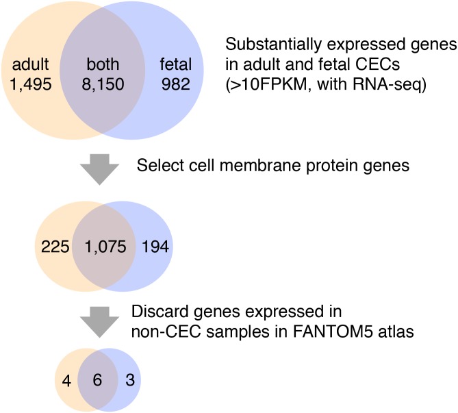
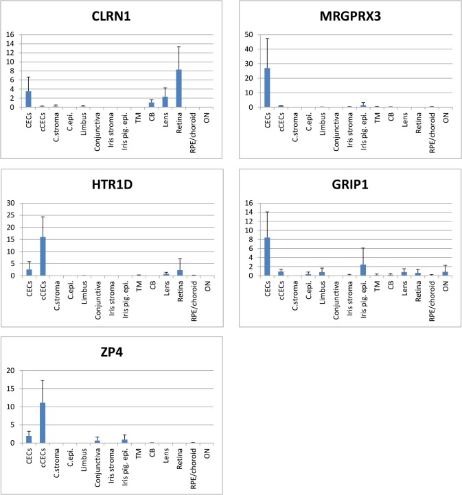
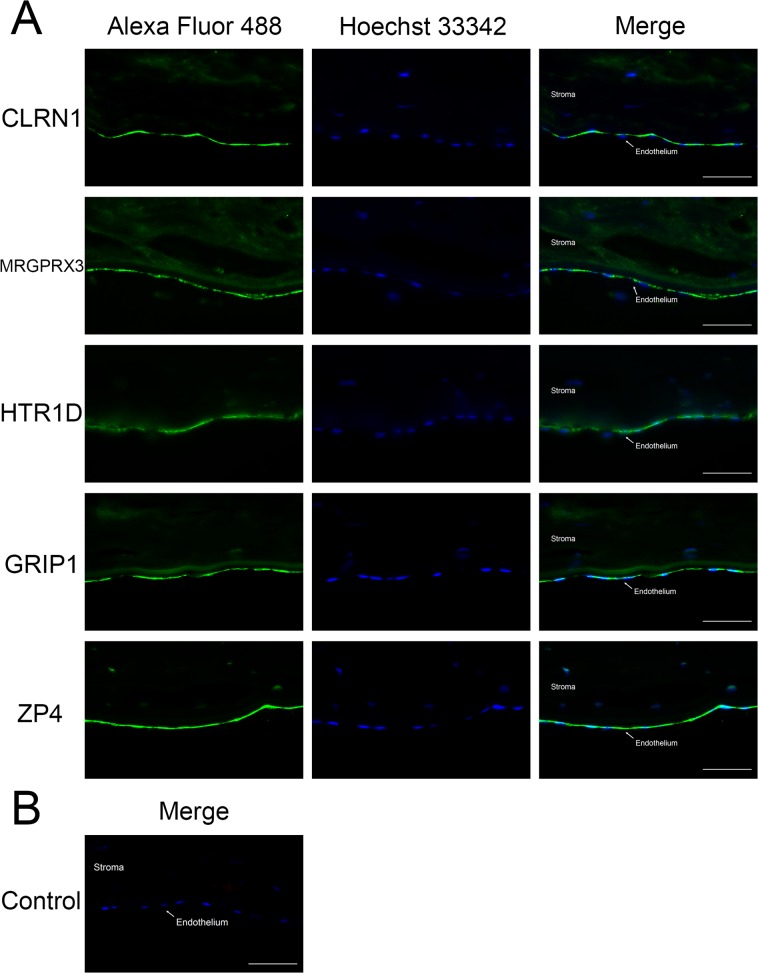
The corneal endothelium is a monolayer of hexagonal corneal endothelial cells (CECs) on the inner surface of the cornea. CECs are critical in maintaining corneal transparency through their barrier and pump functions. CECs in vivo have a limited capacity in proliferation, and loss of a significant number of CECs results in corneal edema called bullous keratopathy which can lead to severe visual loss. Corneal transplantation is the most effective method to treat corneal endothelial dysfunction, where it suffers from donor shortage. Therefore, regeneration of CECs from other cell types attracts increasing interests, and specific markers of CECs are crucial to identify actual CECs. However, the currently used markers are far from satisfactory because of their non-specific expression in other cell types. Here, we explored molecular markers to discriminate CECs from other cell types in the human body by integrating the published RNA-seq data of CECs and the FANTOM5 atlas representing diverse range of cell types based on expression patterns. We identified five genes, CLRN1, MRGPRX3, HTR1D, GRIP1 and ZP4 as novel markers of CECs, and the specificities of these genes were successfully confirmed by independent experiments at both the RNA and protein levels. Notably none of them have been documented in the context of CEC function. These markers could be useful for the purification of actual CECs, and also available for the evaluation of the products derived from other cell types. Our results demonstrate an effective approach to identify molecular markers for CECs and open the door for the regeneration of CECs in vitro.
Conflict of interest statement
Figures
References
-
- Joyce NC, Meklir B, Joyce SJ, Zieske JD (1996) Cell cycle protein expression and proliferative status in human corneal cells. Invest Ophthalmol Vis Sci 37: 645–655. - PubMed
-
- Joyce NC, Navon SE, Roy S, Zieske JD (1996) Expression of cell cycle-associated proteins in human and rabbit corneal endothelium in situ. Invest Ophthalmol Vis Sci 37: 1566–1575. - PubMed
-
- Slingsby JG, Forstot SL (1981) Effect of blunt trauma on the corneal endothelium. Arch Ophthalmol 99: 1041–1043. - PubMed
Publication types
MeSH terms
Substances
Grants and funding
LinkOut - more resources
Full Text Sources
Other Literature Sources
