. 2015 Jun;77(6):1060-75.
doi: 10.1002/ana.24407. Epub 2015 Apr 23.

Voxel-based morphometric magnetic resonance imaging (MRI) postprocessing in MRI-negative epilepsies

Affiliations

Voxel-based morphometric magnetic resonance imaging (MRI) postprocessing in MRI-negative epilepsies

Z Irene Wang et al. Ann Neurol. 2015 Jun.

Abstract

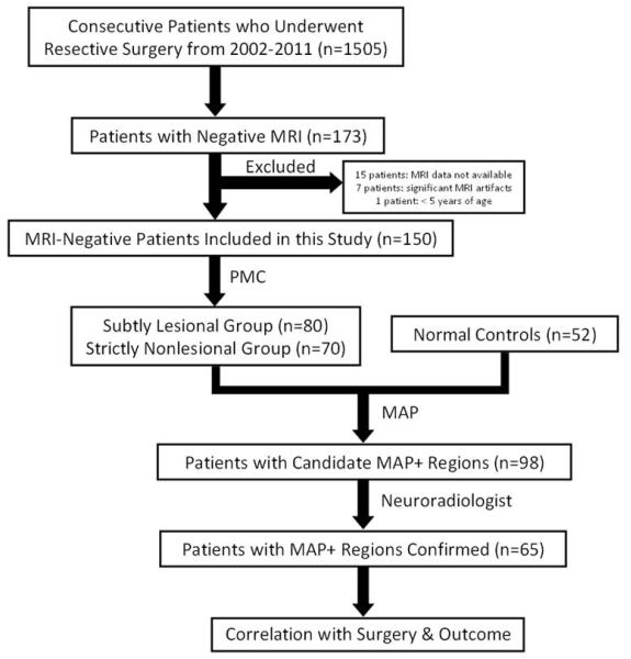
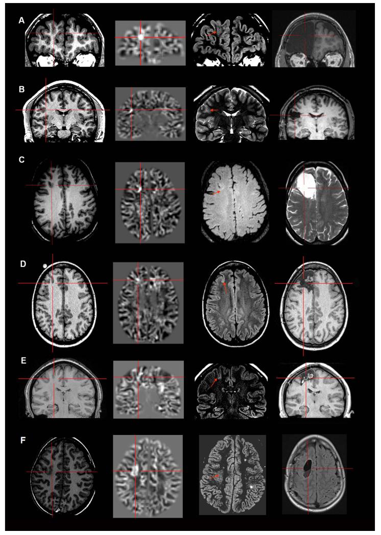
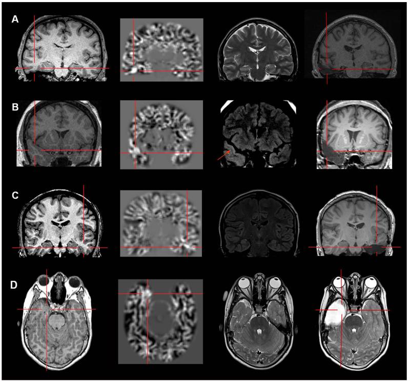
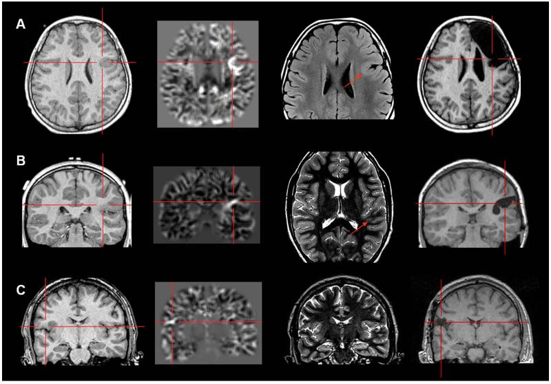
Objective: In the presurgical workup of magnetic resonance imaging (MRI)-negative (MRI(-) or "nonlesional") pharmacoresistant focal epilepsy (PFE) patients, discovering a previously undetected lesion can drastically change the evaluation and likely improve surgical outcome. Our study utilizes a voxel-based MRI postprocessing technique, implemented in a morphometric analysis program (MAP), to facilitate detection of subtle abnormalities in a consecutive cohort of MRI(-) surgical candidates.

Methods: Included in this retrospective study was a consecutive cohort of 150 MRI(-) surgical patients. MAP was performed on T1-weighted MRI, with comparison to a scanner-specific normal database. Review and analysis of MAP were performed blinded to patients' clinical information. The pertinence of MAP(+) areas was confirmed by surgical outcome and pathology.

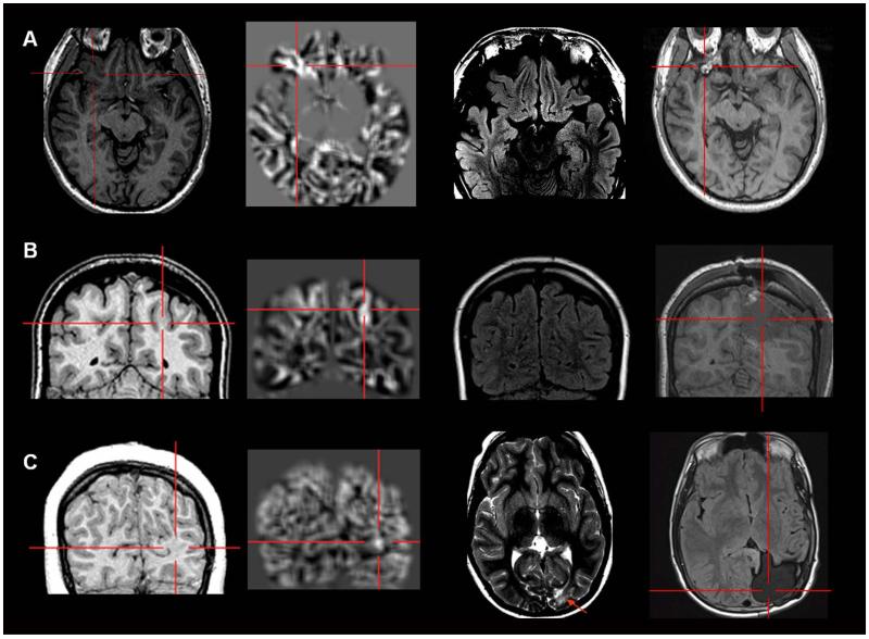
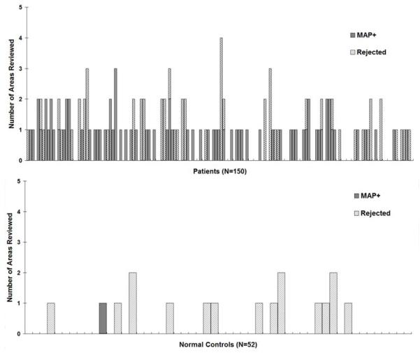
Results: MAP showed a 43% positive rate, sensitivity of 0.9, and specificity of 0.67. Overall, patients with the MAP(+) region completely resected had the best seizure outcomes, followed by the MAP(-) patients, and patients who had no/partial resection of the MAP(+) region had the worst outcome (p < 0.001). Subgroup analysis revealed that visually identified subtle findings are more likely correct if also MAP(+) . False-positive rate in 52 normal controls was 2%. Surgical pathology of the resected MAP(+) areas contained mainly non-balloon-cell focal cortical dysplasia (FCD). Multiple MAP(+) regions were present in 7% of patients.

Interpretation: MAP can be a practical and valuable tool to: (1) guide the search for subtle MRI abnormalities and (2) confirm visually identified questionable abnormalities in patients with PFE due to suspected FCD. A MAP(+) region, when concordant with the patient's electroclinical presentation, should provide a legitimate target for surgical exploration.


Figures

Figure 1
Figure 1
Workflow of data collection and analysis. Patients were retrospectively screened according to the inclusion and exclusion criteria. The included patients were then divided into a “Subtly Lesional” subgroup and a “Strictly Nonlesional” subgroup based on the clinical notes from discussion at the patient management conference (PMC). Normal controls were randomly mixed with the patients. All the patients and controls underwent MAP processing. Candidate MAP+ regions were presented to a neuroradiologist who performed a focused re-review of the original MRI based on the MAP findings. Only when the neuroradiologist agreed that the original MRI showed subtle abnormalities at these sites, the patient was denoted as MAP+. The MAP+ regions were then compared with the location of surgical resection, and association with surgical pathology and seizure outcomes was calculated.
Figure 2
Figure 2
Examples of six patients with MAP+ region in the frontal lobe. In Figures 2-5, the crosshairs pinpoint the location of MAP+ region. First column: T1-weighted MPRAGE images used during pre-surgical evaluation. Second column: gray-white matter junction z-score file, as the output of MAP processing of the T1-weighted image shown in column one. Third column: T2-weighted FLAIR or TSE images, chosen to best depict the MAP+ region. The arrow indicates there was accompanying T2-weighted changes in the MAP+ region, and absence of the arrow indicates that there was no corresponding T2 changes noted. Fourth column: post-surgical MRI indicating site and extent of resection. Panels A and B belong to the “Subtly Lesional” subgroup; C, D, E and F belong to the “Strictly Nonlesional” subgroup. All six cases demonstrate complete resection of the MAP+ region, and all patients remained seizure-free at 12 months. Pathology: A, C, E, F: FCD type I; B, D: FCD type II.
Figure 3
Figure 3
Examples of four patients with MAP+ region in the temporal lobe. Panels A, B and C belong to the “Strictly Nonlesional” subgroup, and Panel D belongs to the “Subtly Lesional” subgroup. All three cases demonstrate complete resection of the MAP+ region, and all patients remained seizure-free at 12 months. Pathology: A, B, C: FCD type I; D: FCD type IIIa.
Figure 4
Figure 4
Examples of three patients with MAP+ region in the operculum (A and C: frontal operculum; B: parietal operculum). Panel A belongs to the “Subtly Lesional” subgroup; B and C belong to the “Strictly Nonlesional” subgroup. All three cases demonstrate complete resection of the MAP+ region, and all patients remained seizure-free at 12 months. Pathology: A, B: FCD type I; C: FCD type II.
Figure 5
Figure 5
Examples of patients with MAP+ region in the orbito-frontal region (A), parietal lobe (B) and occipital lobe (C). Panel A and B belong to the “Strictly Nonlesional” subgroup; panel C belong to the “Subtly Lesional” subgroup. All three cases demonstrate complete resection of the MAP+ region, and all patients remained seizure-free at 12 months. Pathology: A, C: FCD type I; B: FCD type I + nodular heterotopia. In Panel C, the MAP+ region is best shown on the coronal slices; the T2-weighted image and post-operative MRI were only available in axial slices, and therefore only axial slices are presented here.
Figure 6
Figure 6
Detailed information on MAP reviewing, including the number of candidate MAP+ areas reviewed and the number of areas rejected by the neuroradiologist. In both panels, each bar represents one patient/subject. The height of the bars indicates the total number of candidate MAP+ areas reviewed in each patient/subject. Within each bar, the gridded pattern shows the areas reviewed but judged to be non-significant by the neuro-radiologist (and therefore rejected); the solid pattern shows the areas confirmed to be MAP+. For patients/subjects whose MAP did not have any regions with z>4 on the junction file, the height of the bar is set to 0.

References

    1. Bien CG. Characteristics and surgical outcomes of patients with refractory magnetic resonance imaging-negative epilepsies. Arch Neurol. 2009;66:1491–9. - PubMed
    1. Jeha LE. Surgical outcome and prognostic factors of frontal lobe epilepsy surgery. Brain. 2007;130:574–84. - PubMed
    1. Tellez-Zenteno JF, Hernandez Ronquillo L, Moien-Afshari F, Wiebe S. Surgical outcomes in lesional and non-lesional epilepsy: a systematic review and meta-analysis. Epilepsy Res. 2010 May;89(2-3):310–8. - PubMed
    1. Wang ZI, Alexopoulos AV, Jones SE, Jaisani Z, Najm IM, Prayson RA. The pathology of magnetic-resonance-imaging-negative epilepsy. Mod Pathol. 2013 Aug;26(8):1051–8. - PubMed
    1. Hauptman JS, Mathern GW. Surgical treatment of epilepsy associated with cortical dysplasia: 2012 update. Epilepsia. 2012 Sep;53(Suppl 4):98–104. - PubMed

Publication types