Review
. 2015 Aug 20;23(6):565-77.
doi: 10.1089/ars.2015.6306. Epub 2015 Apr 28.

Function and Mechanisms of Autophagy in Brain and Spinal Cord Trauma

Affiliations
Review

Function and Mechanisms of Autophagy in Brain and Spinal Cord Trauma

Marta M Lipinski et al. Antioxid Redox Signal. .

Abstract

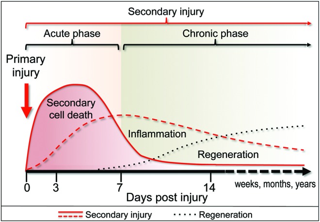
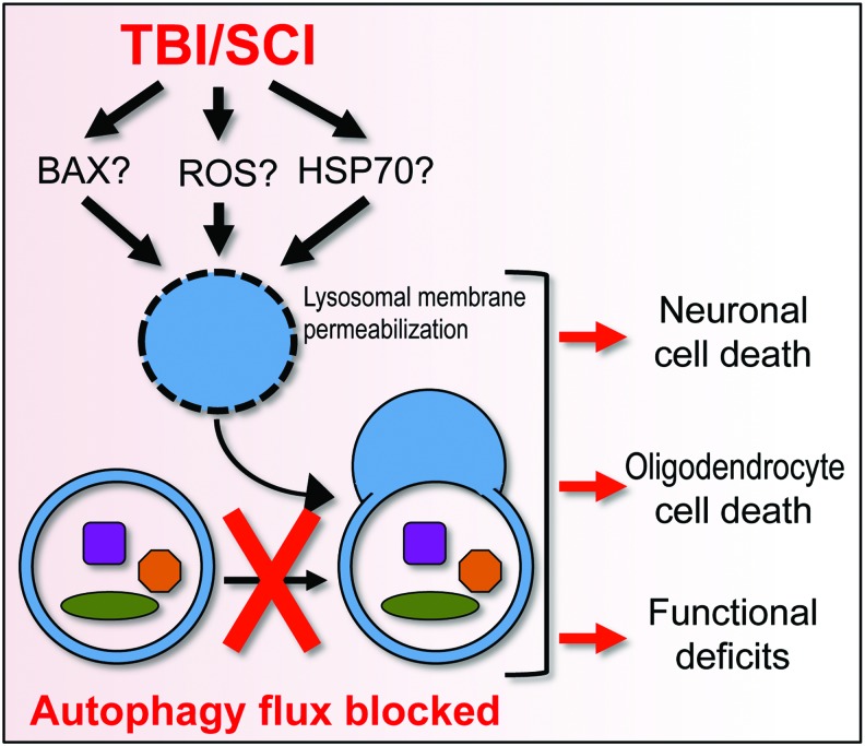
Significance: Traumatic brain injury (TBI) and spinal cord injury (SCI) are major causes of death and long-term disability worldwide. Despite important pathophysiological differences between these disorders, in many respects, mechanisms of injury are similar. During both TBI and SCI, some cells are directly mechanically injured, but more die as a result of injury-induced biochemical changes (secondary injury). Autophagy, a lysosome-dependent cellular degradation pathway with neuroprotective properties, has been implicated both clinically and experimentally in the delayed response to TBI and SCI. However, until recently, its mechanisms and function remained unknown, reflecting in part the difficulty of isolating autophagic processes from ongoing cell death and other cellular events.

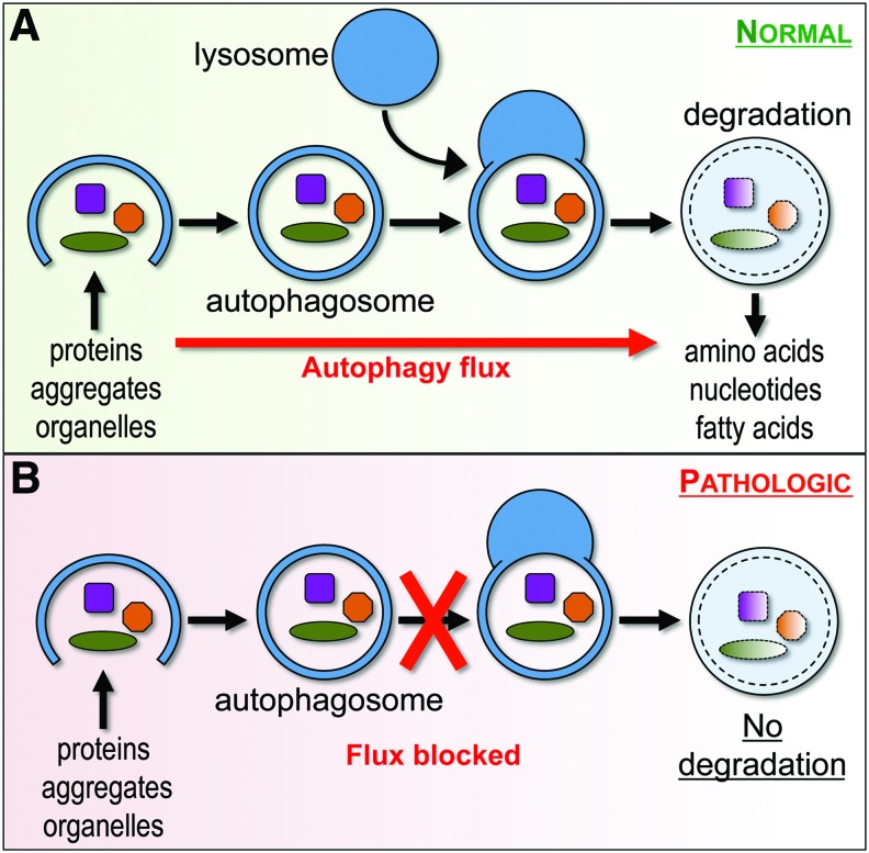
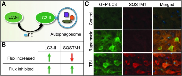
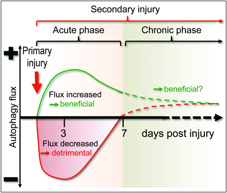
Recent advances: Emerging data suggest that depending on the location and severity of traumatic injury, autophagy flux--defined as the progress of cargo through the autophagy system and leading to its degradation--may be either increased or decreased after central nervous system trauma.

Critical issues: While increased autophagy flux may be protective after mild injury, after more severe trauma inhibition of autophagy flux may contribute to neuronal cell death, indicating disruption of autophagy as a part of the secondary injury mechanism.

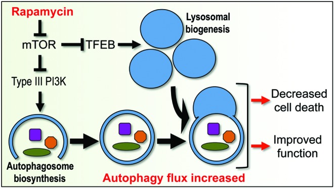
Future directions: Augmentation and/or restoration of autophagy flux may provide a potential therapeutic target for treatment of TBI and SCI. Development of those treatments will require thorough characterization of changes in autophagy flux, its mechanisms and function over time after injury.


Figures

<b>FIG. 1.</b>
FIG. 1.
Sequence of events after CNS trauma in rodent models. The initial mechanical impact (primary injury—red arrow) disrupts the structure of the brain or spinal cord and initiates complex secondary changes that collectively spread the damage to the intact neighboring tissue (secondary injury—red lines). The secondary injury includes secondary neuronal cell death as well as long-term inflammatory changes, which contribute to further damage. Cell damage and death resulting from secondary injury are followed by a restorative phase (regeneration—black dotted line) during which the brain or the spinal cord remodels itself in an attempt to compensate for the tissue damage. CNS, central nervous system. To see this illustration in color, the reader is referred to the web version of this article at www.liebertpub.com/ars
<b>FIG. 2.</b>
FIG. 2.
Autophagy flux under normal and pathological conditions. (A) During autophagy, double-membrane vesicles (autophagosomes) sequester cytoplasmic components, including damaged organelles and toxic protein aggregates, and then fuse with lysosomes to allow degradation of cargo by lysosomal proteases. This progress of cargo through the autophagy system is termed autophagy flux and generally serves a cytoprotective function. (B) Under pathological conditions, autophagy flux may be blocked, for example due to lysosomal defects. This can lead to accumulation of dysfunctional autophagosomes and contribute to cell damage and death. To see this illustration in color, the reader is referred to the web version of this article at www.liebertpub.com/ars
<b>FIG. 3.</b>
FIG. 3.
Common methods used to assess autophagy and autophagy flux. (A) When autophagy is induced, cytosolic LC3-I protein is covalently conjugated to PE to form LC3-II, which translocates to the autophagosomal membrane. Accumulation of LC3-II can be measured as a marker of autophagosome formation. (B, C) Comparison of LC3-II and SQSTM1 levels under conditions when autophagy flux is increased or inhibited. (B) When flux is induced, numbers of autophagosomes and levels of LC3-II increase but levels of autophagy substrates such as SQSTM1 decrease. When autophagy flux is blocked, both numbers of autophagosomes (LC3-II) and autophagy substrates (SQSTM1) increase. (C) Comparison of levels of LC3 (GFP-LC3 fluorescence—green) and SQSTM1 (immunohistochemistry—red) in the cortex of GFP-Lc3 transgenic autophagy reporter mice under conditions when autophagy flux is induced (in vivo Rapamycin treatment for 48 h) versus when autophagy flux is inhibited (24 h after controlled cortical impact injury). Data adapted from Sarkar et al. (53) with permission of authors. PE, phosphatidylethanolamine.
<b>FIG. 4.</b>
FIG. 4.
Changes in autophagy flux after mild versus severe CNS trauma. In some models, TBI can lead to an increase in autophagy flux (green line), which likely serves a protective function. In other models, TBI or SCI can inhibit autophagy flux (red line), contributing to secondary injury. At later time points (dashed lines), autophagy flux may be increased in all models as compared with uninjured animals, suggesting a potential beneficial function. SCI, spinal cord injury; TBI, traumatic brain injury. To see this illustration in color, the reader is referred to the web version of this article at www.liebertpub.com/ars
<b>FIG. 5.</b>
FIG. 5.
Potential mechanisms and consequences of inhibition of autophagy flux after CNS trauma. SCI or severe TBI lead to accumulation of ROS, increased levels of pro-apoptotic BCL2 family proteins, and a decrease in HSP70 chaperone. This can cause lysosomal damage and consequent inhibition of autophagy flux, leading to neuronal and oligodendrocyte cell death and increasing functional deficits after injury. ROS, reactive oxygen species. To see this illustration in color, the reader is referred to the web version of this article at www.liebertpub.com/ars
<b>FIG. 6.</b>
FIG. 6.
Potential mechanisms and consequences of restoring or increasing autophagy flux after CNS trauma. Inhibition of mTOR can increase initiation of autophagy by stimulating activity of the type III PI3 kinase that is necessary for initiation of autophagosome biogenesis. Inhibition of mTOR can also lead to activation of transcription of lysosomal genes by TFEB and increased lysosomal biogenesis. This could restore and/or increase autophagy flux after CNS trauma, leading to neuroprotection and functional improvements. TFEB, transcription factor EB. To see this illustration in color, the reader is referred to the web version of this article at www.liebertpub.com/ars

References

    1. Injury Prevention and Control: Traumatic Brain Injury Center for Disease Control and Prevention. http://cdc.gov/traumaticbraininjury/2014 (Accessed date November/26/2014)
    1. Spinal Cord Injury (SCI): Fact Sheet Center for Disease Control and Prevention. http://cdc.gov/TraumaticBrainInjury/scifacts.html 2014. (Accessed date November/26/2014)
    1. Bains M. and Hall ED. Antioxidant therapies in traumatic brain and spinal cord injury. Biochim Biophys Acta 1822: 675–684, 2012 - PMC - PubMed
    1. Basit F, Cristofanon S, and Fulda S. Obatoclax (GX15-070) triggers necroptosis by promoting the assembly of the necrosome on autophagosomal membranes. Cell Death Differ 20: 1161–1173, 2013 - PMC - PubMed
    1. Beattie MS, Hermann GE, Rogers RC, and Bresnahan JC. Cell death in models of spinal cord injury. Prog Brain Res 137: 37–47, 2002 - PubMed

Publication types

MeSH terms

Substances

LinkOut - more resources