. 2015 Mar 26:6:6646.
doi: 10.1038/ncomms7646.

Mitochondrial protein import receptors in Kinetoplastids reveal convergent evolution over large phylogenetic distances

Affiliations

Mitochondrial protein import receptors in Kinetoplastids reveal convergent evolution over large phylogenetic distances

Jan Mani et al. Nat Commun. .

Abstract

Mitochondrial protein import is essential for all eukaryotes and mediated by hetero-oligomeric protein translocases thought to be conserved within all eukaryotes. We have identified and analysed the function and architecture of the non-conventional outer membrane (OM) protein translocase in the early diverging eukaryote Trypanosoma brucei. It consists of six subunits that show no obvious homology to translocase components of other species. Two subunits are import receptors that have a unique topology and unique protein domains and thus evolved independently of the prototype receptors Tom20 and Tom70. Our study suggests that protein import receptors were recruited to the core of the OM translocase after the divergence of the major eukaryotic supergroups. Moreover, it links the evolutionary history of mitochondrial protein import receptors to the origin of the eukaryotic supergroups.


Figures
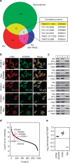
Figure 1
Figure 1. Identification of ATOM complex subunits.
(a) Venn diagram showing the overlap of the T. brucei OM proteome (green) with proteins identified in IPs using mitochondria isolated from cells expressing HA-tagged ATOM40. Elution was either done under denaturing conditions (red) (Supplementary Data 1) or under native condition with subsequent size selection by BN–PAGE (blue) (Supplementary Data 2). (b) Immunofluorescence microscopy of c-Myc-tagged candidate proteins (red) and ATOM40 (green). Merge pictures include staining with 4′,6-diamidino-2-phenylindole (DAPI) to visualize nuclear and mitochondrial DNA (blue). Bar, 10 μm. (c) Immunoblot analysis of c-Myc-tagged candidate proteins in whole cells (T), crude mitochondrial (P) and cytosolic fractions (S). EF1a, mtHSP70 and VDAC served as cytosolic or mitochondrial marker proteins, respectively. (d) Relative abundance of the putative ATOM complex subunits (red) estimated by normalized intensity values of 1,056 proteins identified by mass spectrometry of gradient-purified mitochondria. (e) Relative abundance differences between insect stage (PCF) and bloodstream form (BSF) T. brucei of putative ATOM complex subunits, subunits of the cytochrome c oxidase (COXs) and terminal alternative oxidase (TAO) (see also Supplementary Fig. 1).
Figure 2
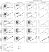
Figure 2. ATOM complex subunits are essential.
Growth curves of uninduced (-Tet) and induced (+Tet) procyclic and bloodstream forms of the indicated knockdown cell lines. Procyclic cells were grown at 27 °C (and for ATOM46 also at 33 °C) and bloodstream forms at 37 °C. All experiments are based on tetracycline (Tet)-inducible RNAi cell lines, except for ATOM11 in procyclic cells for which a conditional knockout cell line was used. This cell line allows depletion of ATOM11 in the absence of Tet. F1γL262P is a bloodstream form cell line that can grow in the absence of the kDNA. It has been tested in the presence of kDNA (positive) or after removal of the kDNA (negative) by treatment with 10 nM ethidiumbromide (Supplementary Fig. 3). Insets, Northern blots or immunoblots confirming successful knockdowns.
Figure 3
Figure 3. In vivo protein import defects.
(a) Immunoblots showing the steady-state levels of ATOM40, VDAC, CoxIV, cytochrome c1 (Cyt c1), MCP5, mtHSP70 and REAP1 in whole-cell extracts of the indicated knockdown cell lines. Cytosolic EF1a serves as a control. Time of induction in days (d) is indicated at the top. Black triangles indicate the onset of the growth phenotype. The position of precursor (p) and mature forms (m) are indicated. (b) Immunoblot analysis of whole-cell (T), digitonin-extracted, mitochondria-enriched pellet (P) and soluble (S) fractions of the indicated knockdown cells. VDAC and EF1a serve as markers for mitochondria and cytosol, respectively. (c) As in a but results are for ATOM69.
Figure 4
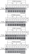
Figure 4. In vitro protein import defects.
35S-Met-labelled LDH–DHFR was imported into mitochondria isolated from the indicated uninduced and induced knockdown cell lines. All import reactions were treated with proteinase K and analysed by SDS–PAGE followed by autoradiography. Ψ, membrane potential. Input, 10% of the added substrate, Coomassie-stained gels are shown as loading controls. The position of precursor (p) and mature forms (m) are indicated.
Figure 5
Figure 5. ATOM complex architecture and functional interactions between its subunits.
(a) BN–PAGE immunoblots of mitochondrial membrane extracts from wild-type cells were probed with antisera against the indicated subunits. For ATOM12, a c-Myc tag version of the protein was analysed. Quantifications of lane profiles revealed the presence of four high molecular weight complexes termed: core, A, B and C. Molecular weight markers (kDa) are indicated. (b) Model of the composition of the complexes. Arrows indicate the suggested assembly pathway. (c) Top panels, SDS–PAGE immunoblot analysis of steady-state levels of individual ATOM complex subunits in all knockdown cell lines. Left and right lanes represent uninduced and induced cell lines, respectively. Lower panel, summary of the immunoblotting data (see also Supplementary Fig. 4).
Figure 6
Figure 6. Domain structure and topology of ATOM complex subunits.
(a) Predicted domain structure of ATOM complex subunits drawn to scale. The predicted domains are indicated in colours. Red, predicted transmembrane domains. (b) Immunoblots of the total (T), pellet (P) and supernatant (S) fractions of carbonate-extracted mitochondria isolated from cells expressing c-Myc-tagged ATOM69 and ATOM46 performed at pH 11.5 and analysed by anti-c-Myc antiserum. VDAC and cytochrome c (Cyt c) serve as marker for an integral and peripheral membrane protein, respectively (c). Immunoblots of a protease protection assay probed for ATOM69 and ATOM46 using gradient-purified wild-type mitochondria. The intermembrane space protein Tim9 serves as a control. Bottom graph, quantification of the ratios between untreated and proteinase K-treated samples of the indicated proteins. (d) Immunoblot analysis of subcellular fractions from transgenic trypansomes expressing c-Myc-tagged variants of ATOM69 and ATOM46 that lack the predicted transmembrane domains. Whole cells (T), digitonin-extracted crude mitochondria (P) and cytosol (S) were analysed. VDAC and EF1a serve as mitochondrial or cytosolic markers, respectively. TEV, TEV protease cleavage site. Asterisks indicate the untagged versions of ATOM69 and ATOM46.
Figure 7
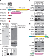
Figure 7. ATOM69 and ATOM46 are novel protein import receptors.
(a) SDS–PAGE of purified His-tagged cytosolic domains of ATOM69 and ATOM46. (b) Binding of a mixture of precursor proteins to the cytosolic domains of ATOM69 and ATOM46. SDS–PAGE analysis showing 10% of the input fractions consisting of the indicated precursor proteins, the eluates from the Ni-NTA beads containing equimolar amounts of the indicated cytosolic domains and, as a control, the eluates from an equal volume of beads only. (c) Immunoblots of steady-state levels of CoxIV and mtHSP70 in whole-cell extracts of the ATOM69 and ATOM46-double knockdown cell line (left panel) or the ATOM46 knockdown cell line (right panel). Cytosolic EF1a serves as a loading control. Time of induction in days (d) is indicated at the top. The black triangle indicates the onset of the growth phenotype. The position of precursor (p) and mature forms (m) are indicated. (d) 35S-Met-labelled LDH-DHFR was imported into mitochondria isolated from the ATOM69 and ATOM46-double knockdown cell line. Ψ, membrane potential. Input, 10% of the added substrate, Coomassie-stained gels are shown as loading controls. The position of precursor (p) and mature forms (m) are indicated.
Figure 8
Figure 8. Diversity of mitochondrial outer membrane protein import receptors.
Three pairs of receptors evolved independently in the Opisthokonta (Tom20/Tom70 shown in blue), in Archeaplastida (Tom20 and OM64 shown in green) and in Kinetoplastids (ATOM46/ATOM69 shown in red). Receptors are drawn to scale and protein domains are indicated. The Stramenopile Tom70 is an orthologue of the Tom70 found in the Opisthokonta, indicating that the Stramenopiles are related to the Opisthokonta or that the Stramenopile Tom70 was acquired by horizontal gene transfer (HGT). ATOM46 and ATOM69 have only been found in Kinetoplastids, indicating that other Excavata lost the receptors maybe due to reductive evolution or that the Excavata are not a monophyletic group.

References

    1. Lithgow T. & Schneider A. Evolution of macromolecular import pathways in mitochondria, hydrogenosomes and mitosomes. Philos. Trans. R. Soc. Lond. B Biol. Sci 365, 799–817 (2010) . - PMC - PubMed
    1. Dolezal P., Likic V., Tachezy J. & Lithgow T. Evolution of the molecular machines for protein import into mitochondria. Science 313, 314–318 (2006) . - PubMed
    1. Hewitt V., Alcock F. & Lithgow T. Minor modifications and major adaptations: the evolution of molecular machines driving mitochondrial protein import. Biochim. Biophys. Acta 1808, 947–954 (2011) . - PubMed
    1. Schmidt O., Pfanner N. & Meisinger C. Mitochondrial protein import: from proteomics to functional mechanisms. Nat. Rev. Mol. Cell Biol. 11, 655–667 (2010) . - PubMed
    1. Neupert W. & Herrmann J. M. Translocation of proteins into mitochondria. Annu. Rev. Biochem. 76, 723–749 (2007) . - PubMed

Publication types

MeSH terms

Substances