

Visualization of pulmonary clearance mechanisms via noninvasive optical imaging validated by near-infrared flow cytometry
- PMID: 25808737
- PMCID: PMC4860906
- DOI: 10.1002/cyto.a.22658
Visualization of pulmonary clearance mechanisms via noninvasive optical imaging validated by near-infrared flow cytometry
Abstract
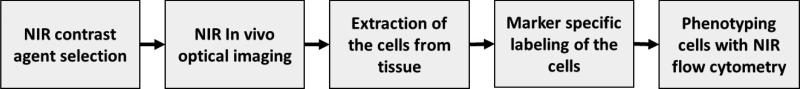
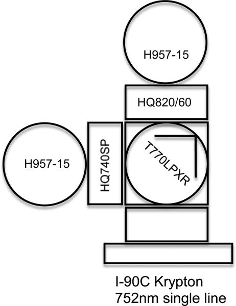
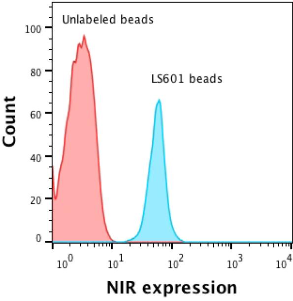
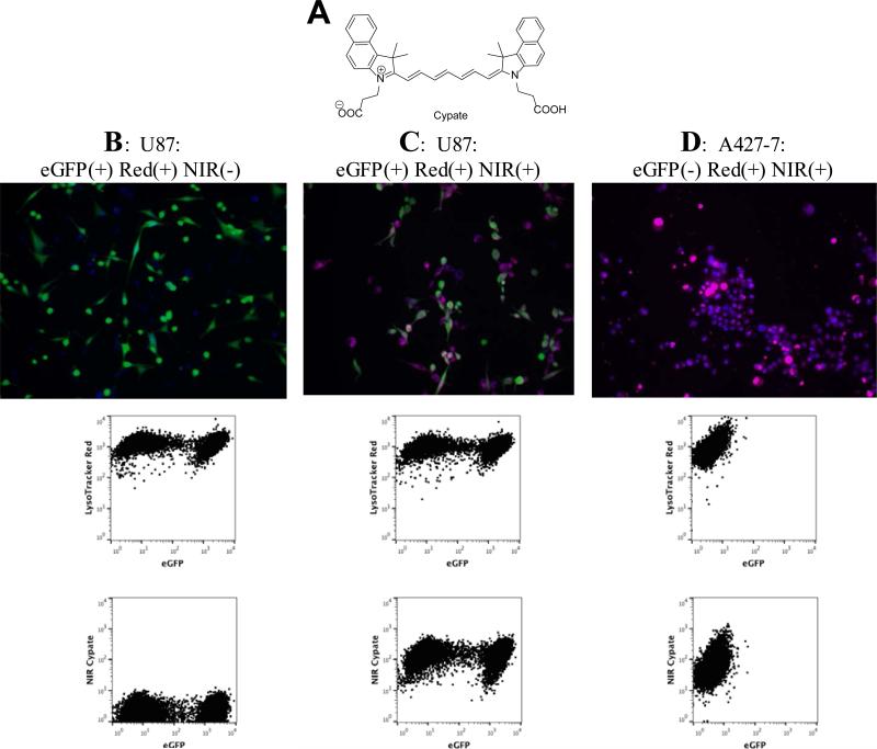
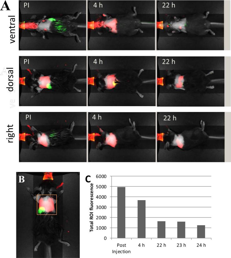
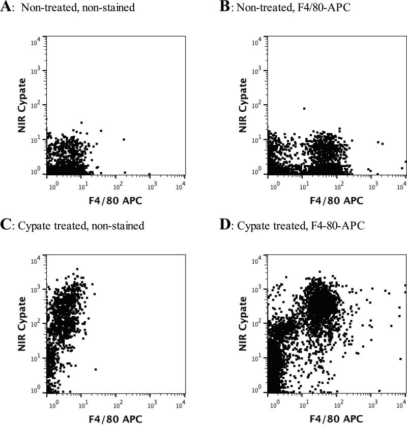
In vivo optical imaging with near-infrared (NIR) probes is an established method of diagnostics in preclinical and clinical studies. However, the specificities of these probes are difficult to validate ex vivo due to the lack of NIR flow cytometry. To address this limitation, we modified a flow cytometer to include an additional NIR channel using a 752 nm laser line. The flow cytometry system was tested using NIR microspheres and cell lines labeled with a combination of visible range and NIR fluorescent dyes. The approach was verified in vivo in mice evaluated for immune response in lungs after intratracheal delivery of the NIR contrast agent. Flow cytometry of cells obtained from the lung bronchoalveolar lavage demonstrated that the NIR dye was taken up by pulmonary macrophages as early as 4-h post-injection. This combination of optical imaging with NIR flow cytometry extends the capability of imaging and enables complementation of in vivo imaging with cell-specific studies.
Keywords: alveolar macrophage; flow cytometry; in vivo imaging; lung imaging; near-infrared.
© 2015 International Society for Advancement of Cytometry.
Figures
References
-
- Ahmed M, Purushotham AD, Douek M. Novel techniques for sentinel lymph node biopsy in breast cancer: a systematic review. Lancet Oncol. 2014;15:e351–e362. - PubMed
-
- Hilderbrand SA, Weissleder R. Near-infrared fluorescence: application to in vivo molecular imaging. Curr Opin Chem Biol. 2010;14:71–9. - PubMed
-
- Weissleder R, Ntziachristos V. Shedding light onto live molecular targets. Nat Med. 2003;9:123–8. - PubMed
-
- Achilefu S, Dorshow RB, Bugaj JE, Rajagopalan R. Novel receptor-targeted fluorescent contrast agents for in vivo tumor imaging. Invest Radiol. 2000;35:479–85. - PubMed
-
- Wessels JT, Busse AC, Mahrt J, Dullin C, Grabbe E, Mueller GA. In vivo imaging in experimental preclinical tumor research--a review. Cytometry A. 2007;71A:542–9. - PubMed
Publication types
MeSH terms
Substances
Grants and funding
LinkOut - more resources
Full Text Sources
Other Literature Sources
Medical
Miscellaneous
