. 2016 May;55(5):671-87.
doi: 10.1002/mc.22312. Epub 2015 Mar 25.

Cathepsin B promotes colorectal tumorigenesis, cell invasion, and metastasis

Affiliations

Cathepsin B promotes colorectal tumorigenesis, cell invasion, and metastasis

Benjamin Bian et al. Mol Carcinog. 2016 May.

Abstract

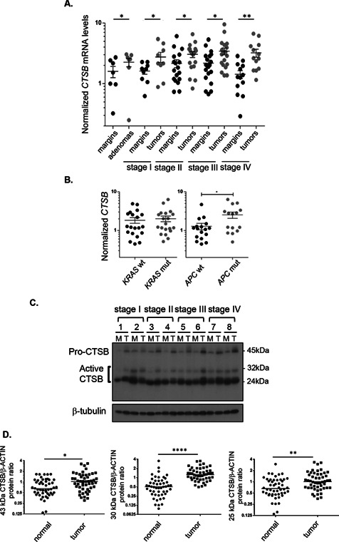
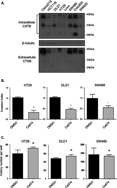
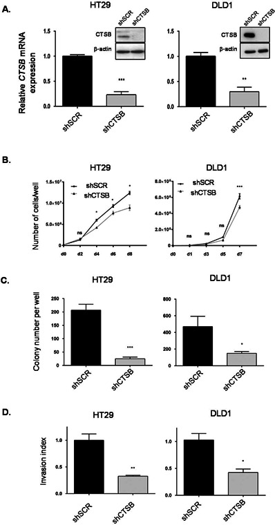
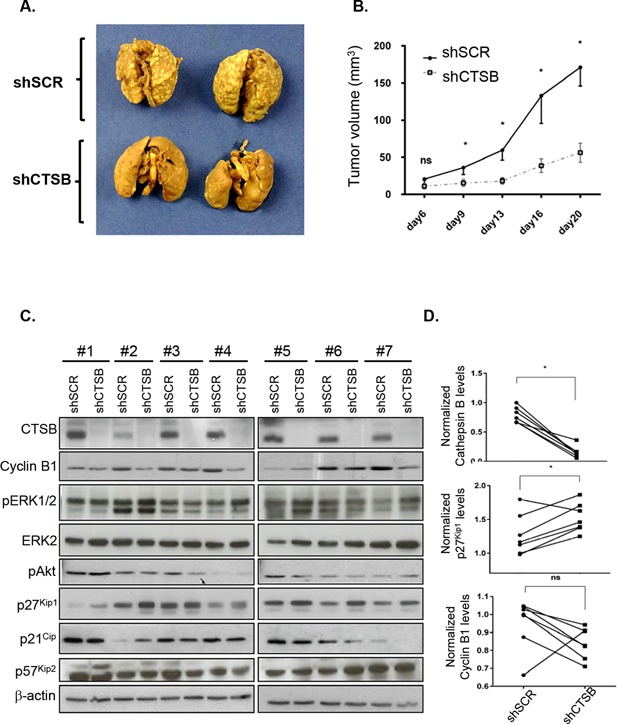
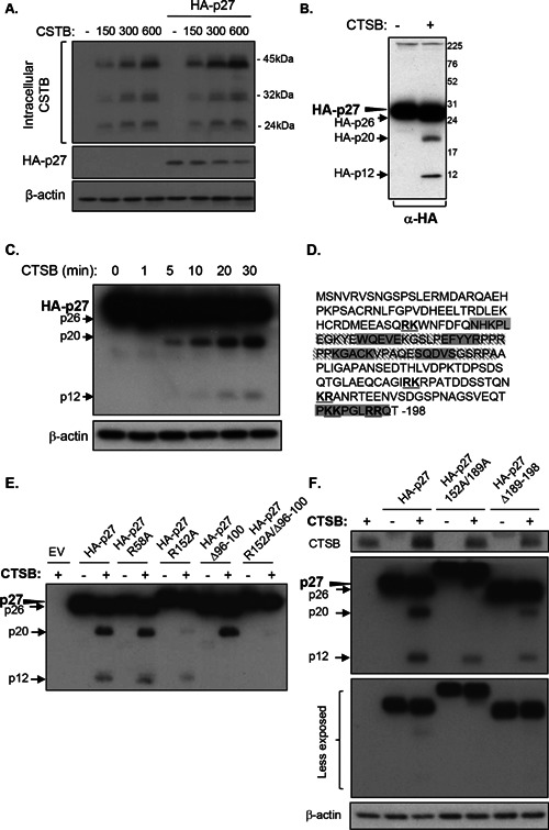
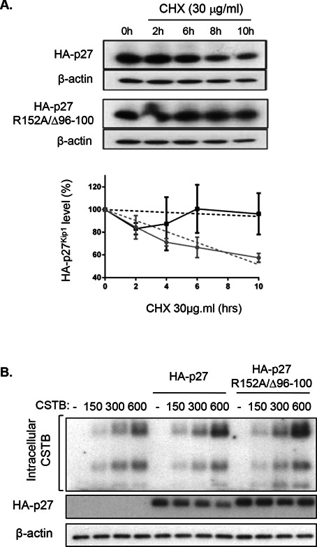
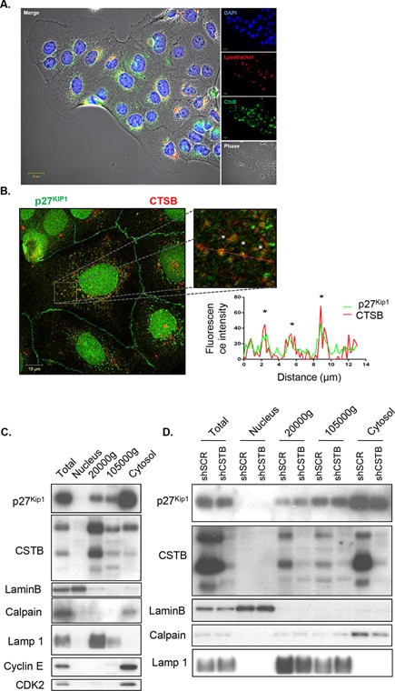
Cathepsin B is a cysteine proteinase that primarily functions as an endopeptidase within endolysosomal compartments in normal cells. However, during tumoral expansion, the regulation of cathepsin B can be altered at multiple levels, thereby resulting in its overexpression and export outside of the cell. This may suggest a possible role of cathepsin B in alterations leading to cancer progression. The aim of this study was to determine the contribution of intracellular and extracellular cathepsin B in growth, tumorigenesis, and invasion of colorectal cancer (CRC) cells. Results show that mRNA and activated levels of cathepsin B were both increased in human adenomas and in CRCs of all stages. Treatment of CRC cells with the highly selective and non-permeant cathepsin B inhibitor Ca074 revealed that extracellular cathepsin B actively contributed to the invasiveness of human CRC cells while not essential for their growth in soft agar. Cathepsin B silencing by RNAi in human CRC cells inhibited their growth in soft agar, as well as their invasion capacity, tumoral expansion, and metastatic spread in immunodeficient mice. Higher levels of the cell cycle inhibitor p27(Kip1) were observed in cathepsin B-deficient tumors as well as an increase in cyclin B1. Finally, cathepsin B colocalized with p27(Kip1) within the lysosomes and efficiently degraded the inhibitor. In conclusion, the present data demonstrate that cathepsin B is a significant factor in colorectal tumor development, invasion, and metastatic spreading and may, therefore, represent a potential pharmacological target for colorectal tumor therapy.

Keywords: cathepsin B; colorectal cancers; intestinal tumorigenesis; invasion; p27Kip1.


Figures

Figure 1
Figure 1
Cathepsin B expression is increased in adenomas and in colorectal cancers of all stages. (A) Relative CTSB mRNA levels were determined by Q‐PCR in human advanced adenomas and adenocarcinomas compared to the paired adjacent healthy tissue. Data presented are the means ± SEM of 7 to 20 tissue samples per stage. Significantly different at *P < 0.05 (Student's t‐test); **P < 0.005 (Student's t‐test). (B) Correlation of induction levels of CTSB transcripts with the presence of KRAS or APC mutations. The induction of CTSB transcripts was analyzed in 15–20 tumors in terms of KRAS and APC mutation status. Data presented are the means ± SEM. Significantly different at *P < 0.05 (Student's t‐test). (C) Representative immunoblot analysis of cathepsin B protein (CTSB) performed on protein extracts from eight paired resection margins and tumor specimens. Tubulin expression is shown as a control of protein loading. (D)Western blot analysis of cathepsin B protein expression in a series of 50 paired specimens (resection margins and primary tumors). Protein levels of cathepsin B pro‐form and active forms were normalized to the intensity of β‐actin staining and to a reference sample, resulting in a dimensionless value. Amounts of normalized cathepsin B proteins in tumor tissues relative to their matched normal samples were analyzed by paired Student's t‐test. Significantly different at *P ≤ 0.05, **P ≤ 0.005, and ****P ≤ 0.0005.
Figure 2
Figure 2
Extracellular cathepsin B contributes to invasiveness of human CRC cells but is dispensable for their growth in soft agar. (A) CRC cell lines were serum‐deprived during 24 h and then lysed in Laemmli buffer and analyzed by Western blotting with specific antibodies against cathepsin B (CTSB) and β‐tubulin. Four milliliter of culture medium from each cell line was concentrated and also analyzed by Western blotting for the expression of extracellular cathepsin B. (B) HT29, DLD1, and SW480 seeded in Matrigel‐coated Transwells were treated with DMSO or Ca074 (10 μM) during 48 h. Thereafter, cells were fixed and stained with DAPI solution for assessment of their capacity to invade Matrigel. The experiments were performed in triplicate and the number of control cells treated with DMSO which had migrated was set at 1. Significantly different at *P ≤ 0.05 and **P ≤ 0.005 (Student's t‐test). (C) HT29, DLD1, and SW480 were cultured in soft agarose in the presence or absence of 10 μM Ca074 for 2–3 wk prior to MTT staining. The number of colonies was determined using ImageJ software. Results are the means ± SEM of at least three independent experiments. ns: not significant (Student's t‐test).
Figure 3
Figure 3
Cathepsin B silencing in human CRC cells inhibits growth in soft agar and invasion capacity. (A) HT29 and DLD1 cells were stably infected with lentiviruses encoding for a control shRNA (scrambled sequence, shSCR) or encoding cathepsin B‐specific shRNAs (shCTSB). Stable cell populations were thereafter lysed and RNA isolated to determine CTSB or PBGD gene expression by Q‐PCR. The relative level of CTSB was normalized to the corresponding PBGD mRNA level. Cell populations were also lysed and protein lysates were analyzed by Western blotting for cathepsin B and β‐actin expression. (B) HT29 and DLD1 cells expressing either shSCR or shCTSB were seeded in a 6‐well plate at 2 × 105 (HT‐29) and 1.2 × 105 (DLD1) cells per well. Cells were harvested and counted after different times. (C) HT29 and DLD1 cells were cultured in soft agarose before MTT staining. The number of colonies was determined using ImageJ software. Results are the means ± SEM of at least three independent experiments. (D) HT29 and DLD1 cell populations were seeded in Matrigel‐coated Transwells and cultured during 48 h. Thereafter, cells were fixed and stained with DAPI solution for assessment of their capacity to invade Matrigel. The experiments were performed in triplicate and the number of control cells (shSCR) which had migrated was set at 1. For all experiments described above: *, significantly different from shSCR cells at P < 0.05 (Student's t‐test). **P < 0.005; ***P < 0.001.
Figure 4
Figure 4
Cathepsin B silencing in human CRC cells inhibits tumorigenicity and metastasis in immunodeficient mice. (A) Representative digital images of mouse lungs 28 d after tail vein injection of 106 HT29 cells expressing either shScrambled (shSCR) or shCathepsin B (shCTSB). (B) HT29 cells expressing either shSCR or shCTSB were injected subcutaneously in immunodeficient mice and the growth of tumors (mm3) over time was measured. The results represent the mean tumor volume obtained from two independent experiments in which at least six mice were injected for each cell line. *, significantly different from shCTSB tumors at P < 0.05 (Student's t‐test). (C) Equal amounts of whole cell lysates from tumors were analyzed by Western blotting for the expression of cathepsin B, cyclin B1, phosphorylated ERK1/2 (pERK1/2), total ERK2, phosphorylated Akt (pAkt), p27Kip1, p21, p57Kip2, and β‐actin. (D) Densitometric analysis of cathepsin B, p27Kip1, and cyclin B1 was determined in each tumor (cells expressing shSCR and cells expressing shCTSB were injected in different flanks of the same mouse) using ImageJ software. *, significantly different from shSCR tumors at P < 0.05 (Wilcoxon matched‐pairs signed‐ranks t‐test).
Figure 5
Figure 5
Cathepsin B exhibits endo‐ and exopeptidase activity against p27Kip1. The pcDNA3 vectors without or with the HA‐tagged wild‐type p27Kip1 cDNA were transiently co‐transfected with 0, 150, 300, or 600 ng of the cDNA encoding cathepsin B in 293T cells. After 48 h, lysates were analyzed by Western blotting for the expression of HA‐27Kip1, cathepsin B, and β‐actin. (B and C) The cDNA encoding HA‐tagged p27Kip1 was transfected transiently in 293T cells. After 48 h, cells were lysed and 15 μg of protein lysate was incubated during 30 min (B) or during 1, 5, 10, 20, and 30 min (C) with purified human cathepsin B (300 ng) at 37°C (pH 6.0). Proteins were then solubilized in Laemmli buffer and analyzed by Western blotting for the expression of HA‐27Kip1 and β‐actin. (D) Amino acid sequence of human p27Kip1; putative cleavage sites of cathepsin B are indicated in bold and underlined. The sequence of amino acids deleted in each p27Kip1 deletion mutant generated is highlighted in grey. (E and F) The cDNA encoding HA‐tagged p27Kip1 and indicated mutants were transfected transiently in 293T cells. After 48 h, cells were lysed and 7.5 μg of protein lysates were incubated during 30 min with purified human cathepsin B (300 ng) at 37°C (pH 6.0). Proteins were then solubilized in Laemmli buffer and analyzed by Western blotting for the expression of HA‐27Kip1, cathepsin B, and β‐actin.
Figure 6
Figure 6
Cathepsin B reduces p27Kip1 stability. (A) The cDNA encoding HA‐tagged wild‐type p27Kip1 and R152A/△96–100 mutant were transiently transfected in 293T cells. Twenty‐four hours following transfection, cells were treated with cycloheximide (CHX, 30 μg/mL) and harvested after 0, 2, 6, 8, or 10 h. Cells were processed and lysed at the same time. Expression of HA‐27Kip1 proteins and β‐actin was analyzed by Western blotting. Representative Western blot analysis is shown in upper panel. The graph illustrates the densitometric analysis of data from three independent experiments. HA‐p27Kip1 expression at 0 h of cycloheximide was set at 100%. Relative HA‐p27Kip1 protein levels were calculated using β‐actin as reference. (B) The pcDNA3 vectors without or with the cDNA encoding HA‐tagged wild‐type p27Kip1 or HA‐tagged R152A/△96–100 mutant were transiently co‐transfected with 0, 150, 300, or 600 ng of the cDNA encoding cathepsin B in 293T cells. After 48 h, protein lysates were analyzed by Western blotting for the expression of HA‐27Kip1, cathepsin B, and β‐actin.
Figure 7
Figure 7
Colocalization of p27 with cathepsin B in lysosomes. (A) Representative confocal microscopy images of Caco‐2/15 CRC cells showing cathepsin B (in green), LysoTracker staining (in red), and DAPI (in blue). (B) Representative confocal microscopy images of Caco‐2/15 CRC cells showing cellular distribution of endogenous p27Kip1 (in green) and cathepsin B (in red) in a double immunofluorescence experiment. Boxed region in the low‐magnification image is enlarged on the right. The graph represents fluorescence profiles along the dashed white line showing areas of colocalization (white asterisks). (C) Caco‐2/15 cell lysates were fractionated by differential centrifugation (see Material and Methods), and equal amounts of proteins from each fraction were analyzed by Western blotting for the expression of p27Kip1 and cathepsin B. Expression of lysosomal (LAMP1), nuclear (lamin B), cytosolic (calpain 2), and nuclear/cytoplasm shuttling markers (cyclin E and CDK2) was analyzed to assess purity of subcellular fractions. (D) SW480 cells stably expressing either shSCR or shCTSB were harvested and lysates were fractionated by differential centrifugation (see Material and Methods). Equal amounts of proteins from each fraction were analyzed by Western blotting for the expression of p27Kip1 and cathepsin B. Expression of lysosomal (LAMP1), nuclear (lamin B), and cytosolic (calpain 2) markers was analyzed to assess purity of subcellular fractions.

Similar articles

Cited by

References

    1. Fearon ER, Vogelstein B. A genetic model for colorectal tumorigenesis. Cell 1990; 61:759–767. - PubMed
    1. DeClerck YA, Mercurio AM, Stack MS, et al. Proteases, extracellular matrix, and cancer: A workshop of the path B study section. Am J Pathol 2004; 16:1131–1139. - PMC - PubMed
    1. Gondi CS, Rao JS. Cathepsin B as a cancer target. Expert Opin Ther Targets 2013; 17:281–291. - PMC - PubMed
    1. Yan S, Sloane BF. Molecular regulation of human cathepsin B: Implication in pathologies. Biol Chem 2003; 384:845–854. - PubMed
    1. Hazen LG, Bleeker FE, Lauritzen B, et al. Comparative localization of cathepsin B protein and activity in colorectal cancer. J Histochem Cytochem 2000; 48:1421–1430. - PubMed

Publication types

MeSH terms