. 2015 May 15;75(10):2109-19.
doi: 10.1158/0008-5472.CAN-14-3122. Epub 2015 Mar 25.

Development of Resistance to EGFR-Targeted Therapy in Malignant Glioma Can Occur through EGFR-Dependent and -Independent Mechanisms

Affiliations

Development of Resistance to EGFR-Targeted Therapy in Malignant Glioma Can Occur through EGFR-Dependent and -Independent Mechanisms

Stefan Klingler et al. Cancer Res. .

Abstract

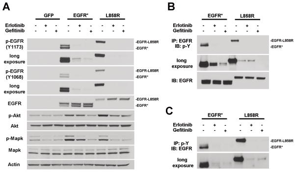
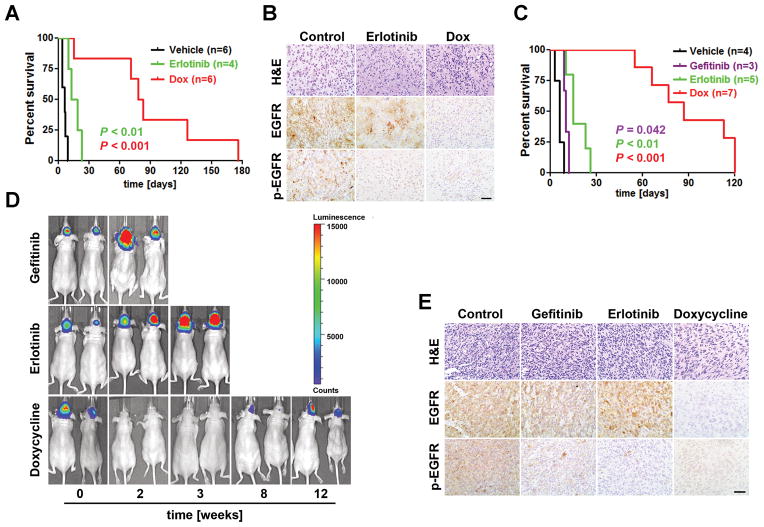
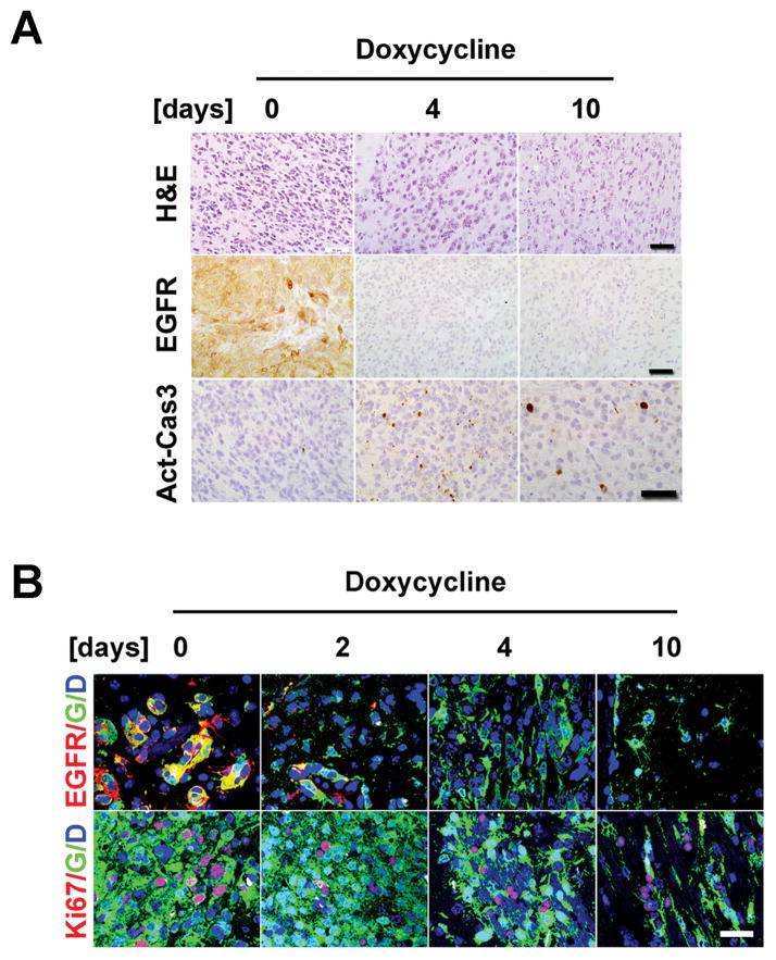
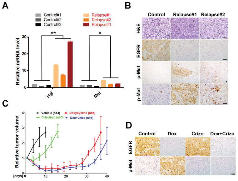
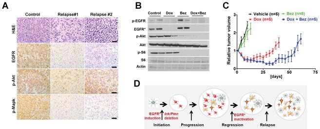
Epidermal growth factor receptor (EGFR) is highly amplified, mutated, and overexpressed in human malignant gliomas. Despite its prevalence and growth-promoting functions, therapeutic strategies to inhibit EGFR kinase activity have not been translated into profound beneficial effects in glioma clinical trials. To determine the roles of oncogenic EGFR signaling in gliomagenesis and tumor maintenance, we generated a novel glioma mouse model driven by inducible expression of a mutant EGFR (EGFR*). Using combined genetic and pharmacologic interventions, we revealed that EGFR*-driven gliomas were insensitive to EGFR tyrosine kinase inhibitors, although they could efficiently inhibit EGFR* autophosphorylation in vitro and in vivo. This is in contrast with the genetic suppression of EGFR* induction that led to significant tumor regression and prolonged animal survival. However, despite their initial response to genetic EGFR* extinction, all tumors would relapse and propagate independent of EGFR*. We further showed that EGFR*-independent tumor cells existed prior to treatment and were responsible for relapse following genetic EGFR* suppression. And, the addition of a PI3K/mTOR inhibitor could significantly delay relapse and prolong animal survival. Our findings shed mechanistic insight into EGFR drug resistance in glioma and provide a platform to test therapies targeting aberrant EGFR signaling in this setting.


Figures

Figure 1
Figure 1
Auto-phosphorylation of EGFR* and EGFR-L858R is efficiently inhibited by EGFR TKIs. A, murine Ink4a/Arf−/− Pten −/− astrocytes transduced with control or indicated EGFR mutants were serum starved for 24 hours followed by 4-hour treatment of vehicle (DMSO), 250 nM erlotinib or 50 nM gefitinib. Cell lysates were prepared and subjected to immunoblot analysis. Note, the molecular weight of EGFR (wt) and EGFR-L858R is ~170KD and EGFR* ~140 KD. B and C, cell lysates from the indicated treatments as in (A) were immunoprecipitated with anti-EGFR (B) or anti-phospho-tyrosine antibody (PT-66) (C), and immunoblot analysis was performed using anti-phospho-tyrosine (4G10) (B) or anti-EGFR (C) antibody, respectively.
Figure 2
Figure 2
EGFR* induction cooperates with conditional Ink4a/Arf and Pten deletions to induce malignant glioma formation. A, schematic of inducible EGFR* allele under regulation of human GFAP promoter element. B, EGFR* induction was efficiently repressed by Dox administration. Brain tissue lysates were prepared from littermate single- or bi-transgenic mice maintained on- (+) or off- (−) Dox and subjected to immunoblot analysis for EGFR and Actin. C, Kaplan-Meier brain tumor-free survival analysis of tamoxifen-treated mouse cohorts consisting of tetO-EGFR* cInkL cPtenL (iEIP) maintained off-Dox (n = 25), tetO-EGFR* cInkL cPtenL (iEIP) on-Dox (n = 5), tetO-EGFR* cInkL (iEI) off-Dox (n = 6), tetO-EGFR* cPtenL (iEP) off-Dox (n = 5), tetO-EGFR* (iEGFR) off-Dox (n = 7), and cInkL cPtenL (cIP) off-Dox (n = 8). D, iEIP gliomas displayed high mitotic indices and low levels of apoptosis. H&E and IHC staining against Ki67 and activated Caspase-3 (Act-Cas3) were performed using sections from normal mouse brains or iEIP malignant gliomas. E, sections of normal mouse brain tissue or iEIP tumors were stained with antibodies against Pten and EGFR. The arrow heads point to embedded Pten-positive endothelial cells in the tumors. F, shown are representative images of IHC staining against phosphorylated Akt (p-Akt), Mapk (p-Mapk) and Stat3 (p-Stat3) performed on sections from normal mouse brains and iEIP malignant gliomas. Scale bars represent 50 μm.
Figure 3
Figure 3
The iEIP malignant gliomas are heterogeneous. A, iEIP glioma cells display multi-lineage differentiation. Normal mouse brain and iEIP glioma sections were stained with H&E or antibodies against indicated lineage markers. B, IHC staining against EGFR was performed on sections from different regions of three independent iEIP gliomas. Stronger EGFR expression was found at invasive edges of tumor periphery compared to relatively solid tumor centers. Scale bars represent 50 μm. C, shown are representative IF images of iEIP gliomas with interspersed EGFR-high and -low tumor cells. Tumor cells were distinguished by their negative Pten expression compared to embedded Pten-positive normal cells (green). D, co-staining of EGFR and Ki67 antibodies revealed that EGFR-high and EGFR-low tumor cells both retained proliferation capacity. Scale bars represent 100 μm.
Figure 4
Figure 4
The iEIP gliomas are sensitive to genetic suppression of EGFR* induction but are refractory to EGFR TKI treatment. A, Kaplan-Meier survival analysis of cohorts of tumor bearing iEIP transgenic mice treated with vehicle (n = 6), erlotinib (n = 4), or Dox (n = 6). Day 0 represents the day when treatment was initiated. B, EGFR* phosphorylation but not EGFR* protein was downregulated in erlotinib treated tumors. Shown are representative images of H&E and IHC staining against EGFR and phospho-EGFR (p-EGFR) on tumor sections from (A). C, Kaplan-Meier survival analysis of mouse cohorts that were orthotopically transplanted with iEIP glioma cells and treated with vehicle (n = 4), gefitinib (n = 3), erlotinib (n = 5), or Dox (n = 7) after tumors were established. Day 0 represents the day when treatment was initiated. D, shown are representative bioluminescence images of animals subjected to the indicated treatment from (C). E, EGFR* phosphorylation but not EGFR* protein levels were diminished in tumors treated with gefitinib or erlotinib. Representative tumor sections from (C) were stained with H&E, anti-EGFR, or anti-p-EGFR. Scale bars represent 50 μm.
Figure 5
Figure 5
EGFR*-independent glioma cells exist prior to treatment. A, tumor bearing animals grafted with GFP-expressing iEIP glioma cells were switched to Dox and sacrificed at indicated time-points (n = 2 for each). H&E and IHC staining against EGFR or activated caspase3 (Act-Cas3) revealed complete suppression of EGFR* induction at 4-day post treatment and enhanced apoptosis in Dox treated tumors. Scale bars represent 50 μm. B, IF staining revealed a subpopulation of GFP-labeled Ki67-positive proliferative tumor cells persisted through genetic suppression of EGFR* induction. G - GFP; D - DAPI. Scale bars represent 100 μm.
Figure 6
Figure 6
Hgf/Met signaling is activated in EGFR*-independent relapsed tumors. A, Total RNAs were prepared from untreated control and Dox-treated relapsed tumors and subjected to qPCR analysis for Hgf, Met and β-Actin. Results were normalized with β-Actin expression and shown as mean ± SD. Student’s t test was used for the comparison between untreated control and Dox-treated relapsed group (**, p = 0.034; *, p = 0.046). Data were from two independent experiments with triplicates. B, Met activated tumor cells were focally distributed in Dox-treated relapsed tumors. Shown are representative images of untreated control and relapsed tumor sections stained for H&E, EGFR and phospho-Met (p-Met). C and D, Met inhibitor had limited effect on iEIP tumor growth and relapse prevention. Mice with subcutaneously grafted iEIP glioma cells were treated with vehicle (n = 4), crizotinib (n = 5), Dox (n = 4), or Dox + crizotinib (n = 5). Day 0 represents the day when treatment was initiated. Tumor growth was measured at indicated time and calculated relative to initial tumor volume. The data are presented as mean ± SD.
Figure 7
Figure 7
Combining PI3K/mTOR inhibition with genetic EGFR* suppression prolongs animal survival by delaying tumor relapse. A, Akt but not Mapk phosphorylation was maintained in Dox-treated relapsed tumors. Shown are representative images of untreated control and relapsed tumor sections stained with antibodies against phospho-Akt (p-Akt) and phospho-Mapk (p-Mapk). Scale bars represent 50 μm. B and C, mice with subcutaneously grafted iEIP glioma cells were treated with vehicle (n = 5), Bez-235 (n = 5), Dox (n = 4), or Dox + Bez-235 (n = 5). Day 0 represents the day when treatment was initiated. Immunoblot analysis (B) was performed using tumor lysates prepared from the indicated treatments. Tumor growth (C) was measured at indicated time and calculated relative to initial tumor volume. The data are presented as mean ± SD. D, model of EGFR*-induced glioma initiation/progression, and its relation to EGFR-targeted therapeutic resistance development.

References

    1. Louis DN, Ohgaki H, Wiestler OD, Cavenee WK, Burger PC, Jouvet A, et al. The 2007 WHO classification of tumours of the central nervous system. Acta Neuropathol. 2007;114:97–109. - PMC - PubMed
    1. Wen PY, Kesari S. Malignant gliomas in adults. N Engl J Med. 2008;359:492–507. - PubMed
    1. Stupp R, Mason WP, van den Bent MJ, Weller M, Fisher B, Taphoorn MJ, et al. Radiotherapy plus concomitant and adjuvant temozolomide for glioblastoma. N Engl J Med. 2005;352:987–96. - PubMed
    1. Brennan CW, Verhaak RG, McKenna A, Campos B, Noushmehr H, Salama SR, et al. The somatic genomic landscape of glioblastoma. Cell. 2013;155:462–77. - PMC - PubMed
    1. Parsons DW, Jones S, Zhang X, Lin JC, Leary RJ, Angenendt P, et al. An integrated genomic analysis of human glioblastoma multiforme. Science. 2008;321:1807–12. - PMC - PubMed

Publication types

MeSH terms