. 2015 Mar 19;7(1):503-37.
doi: 10.3390/cancers7010503.

Stat5 Exerts Distinct, Vital Functions in the Cytoplasm and Nucleus of Bcr-Abl+ K562 and Jak2(V617F)+ HEL Leukemia Cells

Affiliations

Stat5 Exerts Distinct, Vital Functions in the Cytoplasm and Nucleus of Bcr-Abl+ K562 and Jak2(V617F)+ HEL Leukemia Cells

Axel Weber et al. Cancers (Basel). .

Erratum in

Abstract

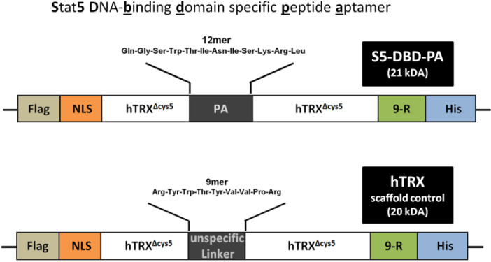
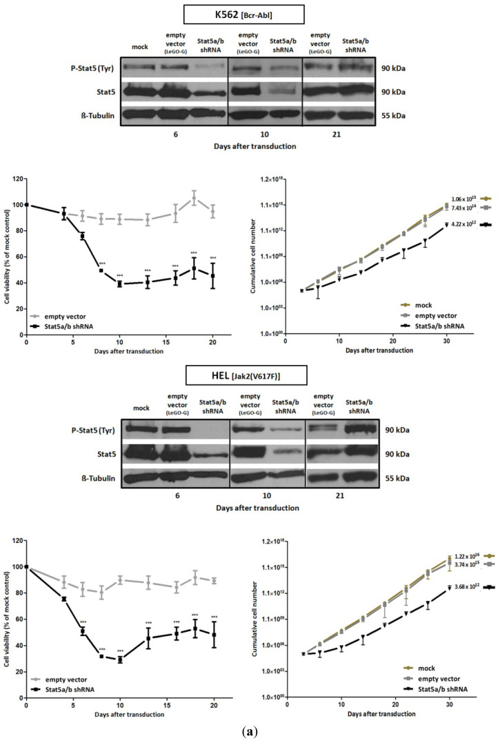
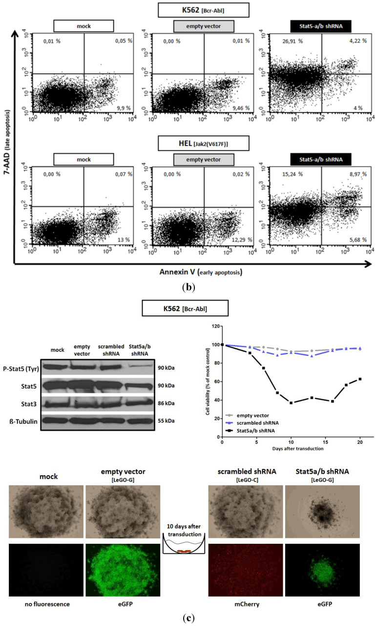
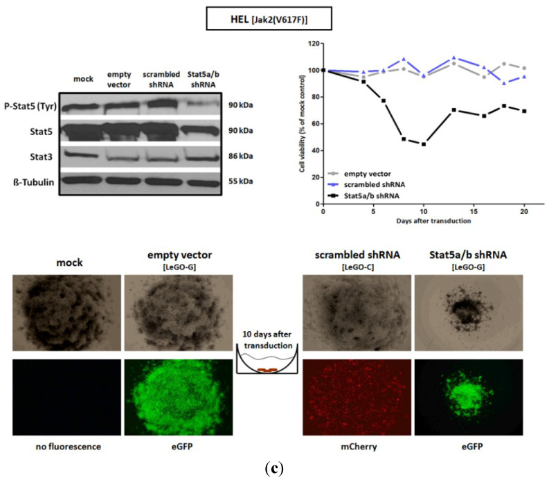
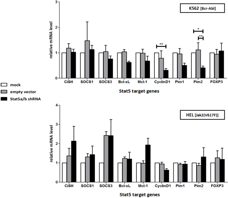
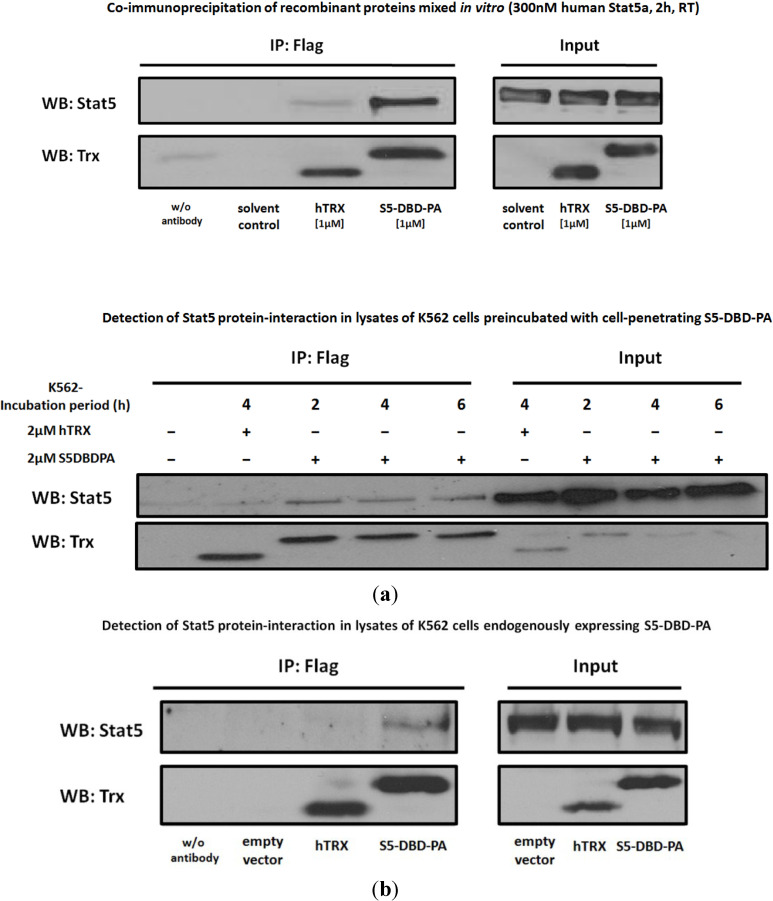
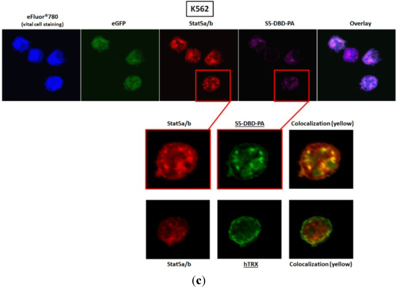
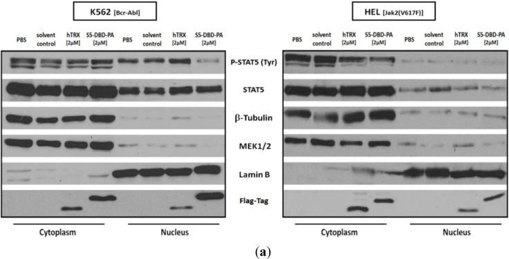
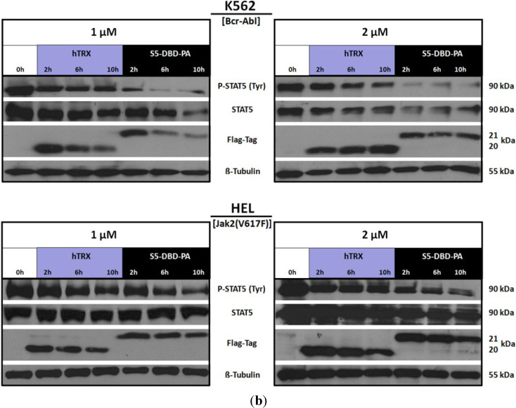
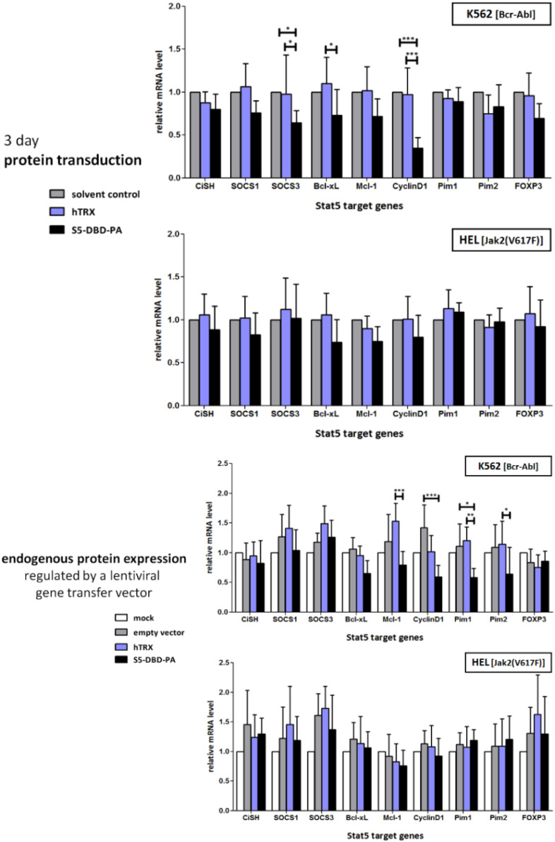
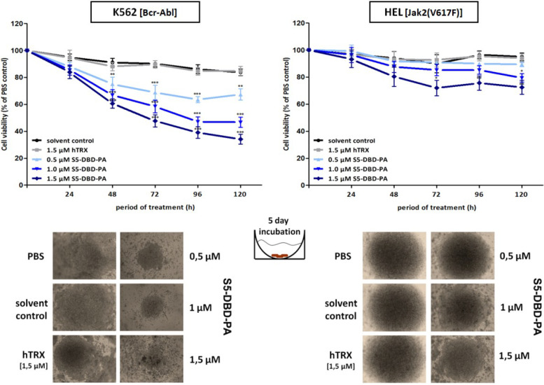
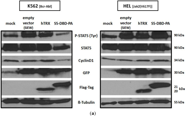
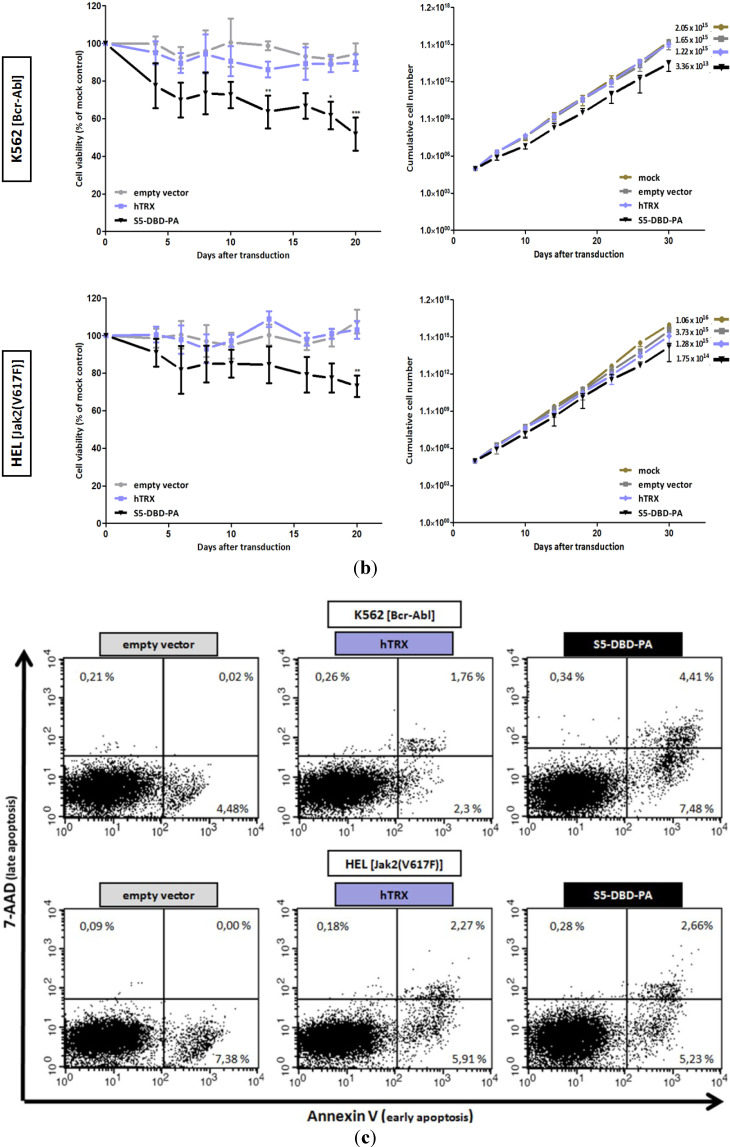
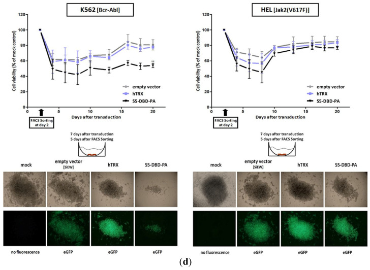
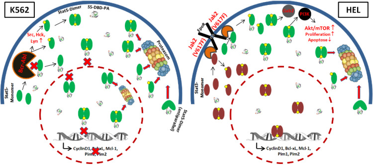
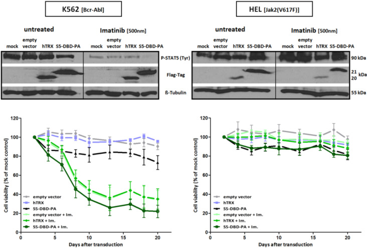
Signal transducers and activators of transcription (Stats) play central roles in the conversion of extracellular signals, e.g., cytokines, hormones and growth factors, into tissue and cell type specific gene expression patterns. In normal cells, their signaling potential is strictly limited in extent and duration. The persistent activation of Stat3 or Stat5 is found in many human tumor cells and contributes to their growth and survival. Stat5 activation plays a pivotal role in nearly all hematological malignancies and occurs downstream of oncogenic kinases, e.g., Bcr-Abl in chronic myeloid leukemias (CML) and Jak2(V617F) in other myeloproliferative diseases (MPD). We defined the mechanisms through which Stat5 affects growth and survival of K562 cells, representative of Bcr-Abl positive CML, and HEL cells, representative for Jak2(V617F) positive acute erythroid leukemia. In our experiments we suppressed the protein expression levels of Stat5a and Stat5b through shRNA mediated downregulation and demonstrated the dependence of cell survival on the presence of Stat5. Alternatively, we interfered with the functional capacities of the Stat5 protein through the interaction with a Stat5 specific peptide ligand. This ligand is a Stat5 specific peptide aptamer construct which comprises a 12mer peptide integrated into a modified thioredoxin scaffold, S5-DBD-PA. The peptide sequence specifically recognizes the DNA binding domain (DBD) of Stat5. Complex formation of S5-DBD-PA with Stat5 causes a strong reduction of P-Stat5 in the nuclear fraction of Bcr-Abl-transformed K562 cells and a suppression of Stat5 target genes. Distinct Stat5 mediated survival mechanisms were detected in K562 and Jak2(V617F)-transformed HEL cells. Stat5 is activated in the nuclear and cytosolic compartments of K562 cells and the S5-DBD-PA inhibitor most likely affects the viability of Bcr-Abl+ K562 cells through the inhibition of canonical Stat5 induced target gene transcription. In HEL cells, Stat5 is predominantly present in the cytoplasm and the survival of the Jak2(V617F)+ HEL cells is impeded through the inhibition of the cytoplasmic functions of Stat5.


Figures

Figure 1
Figure 1
Design of the Stat5 specific peptide aptamer (PA) construct, S5-DBD-PA, a ligand of the DNA-binding domain of Stat5. The recombinant S5-DBD-PA protein comprise functional domains for its purification and transduction into target cells. The 12mer PA sequence, a specific interactor of the DNA-binding domain (DBD) of Stat5, is integrated in the active loop of the optimized hTRXΔcys5 scaffold protein, this provides for a constrained conformation of the peptide favorable for target binding and stability. A domain of 9 sequential L-arginine residues (9-R) serves as a protein transduction domain (PTD) and a histidine tag (His) for purification by nickel-affinity chromatography. These domains are present at the C-terminus of hTRXΔcys5. An N-terminal Flag epitope provides for the immunodetection of the protein and a NLS-sequence allows for enhanced nuclear import. The S5-DBD-PA construct encodes a protein of 21 kDa. The replacement of the 12mer PA sequence by an unspecific 9mer linker sequence yielded a scaffold control protein construct (hTRX, 20 kDa).
Figure 2
Figure 2
shRNA-mediated downregulation of Stat5 expression strongly reduces the survival of Bcr-Abl expressing K562 and Jak2(V617F) expressing HEL cells. (a) shRNA mediated downregulation of Stat5a and Stat5b expression. K562 and HEL cells were transduced with lentiviral vectors encoding shRNA directed against both Stat5 isoforms. Mock treated and empty vector (LeGO-G) transduced cells served as controls. Western blot analyses was used to visualize the expression of Stat5 and activated P-Stat5 6, 10 and 21 days after infection of the cells. Cell proliferation and viability were assayed over a period of 20 days after infection by XTT measurement (n = 4; Ø ± SD). Significantly reduced XTT-values (percentage of mock control) were found when the cells were compared to empty vector expressing cells *** p < 0.001 (2-way-ANOVA with Bonferroni correction). Growth analyses were carried out by counting the cumulative cell numbers at each passage from day 3 to day 30 after infection (n = 3; Ø ± SD); (b) Apoptosis measurement by Annexin V/7-AAD staining. Cells were stained and analyzed 10 days after transduction with shRNA-encoding lentiviral vectors. Divided FACS dot plots indicate unstained vital cells (lower left), early apoptotic cells positive for Annexin V (lower right), Annexin V/7-AAD double positive apoptotic cells (upper right) and late apoptotic/necrotic cells positive for -AAD (upper left); (c) In a control experiment K562 and HEL cells were treated with a lentiviral vector (LeGO-C) expressing a scrambled shRNA. Cell viability was measured over a period of 20 days by XTT conversion, whereas the corresponding suspension cell mass was documented after 10 days in assay-round bottom wells by phase contrast and fluorescence microscopy. After 14 days cell lysates were analyzed by western blotting with antibodies detecting Stat5 or P-Stat5, detection of Stat3 served as a control for the specificity of the of shRNA.
Figure 2
Figure 2
shRNA-mediated downregulation of Stat5 expression strongly reduces the survival of Bcr-Abl expressing K562 and Jak2(V617F) expressing HEL cells. (a) shRNA mediated downregulation of Stat5a and Stat5b expression. K562 and HEL cells were transduced with lentiviral vectors encoding shRNA directed against both Stat5 isoforms. Mock treated and empty vector (LeGO-G) transduced cells served as controls. Western blot analyses was used to visualize the expression of Stat5 and activated P-Stat5 6, 10 and 21 days after infection of the cells. Cell proliferation and viability were assayed over a period of 20 days after infection by XTT measurement (n = 4; Ø ± SD). Significantly reduced XTT-values (percentage of mock control) were found when the cells were compared to empty vector expressing cells *** p < 0.001 (2-way-ANOVA with Bonferroni correction). Growth analyses were carried out by counting the cumulative cell numbers at each passage from day 3 to day 30 after infection (n = 3; Ø ± SD); (b) Apoptosis measurement by Annexin V/7-AAD staining. Cells were stained and analyzed 10 days after transduction with shRNA-encoding lentiviral vectors. Divided FACS dot plots indicate unstained vital cells (lower left), early apoptotic cells positive for Annexin V (lower right), Annexin V/7-AAD double positive apoptotic cells (upper right) and late apoptotic/necrotic cells positive for -AAD (upper left); (c) In a control experiment K562 and HEL cells were treated with a lentiviral vector (LeGO-C) expressing a scrambled shRNA. Cell viability was measured over a period of 20 days by XTT conversion, whereas the corresponding suspension cell mass was documented after 10 days in assay-round bottom wells by phase contrast and fluorescence microscopy. After 14 days cell lysates were analyzed by western blotting with antibodies detecting Stat5 or P-Stat5, detection of Stat3 served as a control for the specificity of the of shRNA.
Figure 3
Figure 3
Stat5 specific shRNA reduces the expression of Stat5 target genes Bcl-xL, Cyclin D1 and Pim2 in Bcr-Abl+ K562, but not in Jak2(V617F)+ HEL cells. Relative expression level of selected Stat5 target genes were analyzed by qRT-PCR in lentivirus transduced K562 and HEL cells. The cells were lysed after seven days and total RNA was extracted for qRT-PCR measurement. Data were normalized to HPRT1 housekeeping gene expression and the relative levels are shown as folds of mock treated control cells (n = 3; Ø ± SD). Significantly reduced gene expression level of shRNA expressing cells in comparison to mock and empty vector expressing control cells are indicated. * p < 0.05, ** p < 0.01 (2-way-ANOVA with Bonferroni correction).
Figure 4
Figure 4
S5-DBD-PA specifically interacts with Stat5 and intracellularly colocalizes with Stat5. (a) Co-immunoprecipitation (Co-IP) of recombinant S5-DBD-PA and Stat5. Co-IP studies were carried out either with a mixture of recombinant S5-DBD-PA (f.c.: 1 µM) and recombinant human Stat5a protein (f.c.: 300 nM), incubated for 2 h at room temperature (RT), or with lysates of K562 cells incubated for 2, 4 and 6 h with S5-DBD-PA (f.c.: 2 µM). S5-DBD-PA was precipitated with a Flag-tag antibody. Immunoprecipitates were examined by Western blotting with thioredoxin and Stat5 specific antibodies. The use of non-antibody loaded beads as well as the incubation with protein solvent (dialysis buffer) and with the non-specific scaffold control protein hTRX (f.c.: 1 or 2 µM) served as controls. During the time of S5-DBD-PA addition to K562 cells, the cells were cultured in starvation medium (2% FCS). Spuriously attached proteins were removed by acid-wash prior to lysate preparation. Input represents 10% of the sample volume used for the Co-IP experiment; (b) Co-IP analysis of the S5-DBD-PA/Stat5 interaction after endogenous expression of S5-DBD-PA in target cells. K562 cells were infected with lentivirus encoding either the empty vector (SiEW) or encoding the S5-DBD-PA and hTRX proteins. Lysates were prepared seven days after infection and used for IP with a Flag-tag antibody. Protein constructs and co-precipitated Stat5 were subsequently analyzed by western blotting with Stat5 or thioredoxin specific antibodies. Unspecific binding was controlled by lysate incubation with beads without antibodies. Input represents 10% of the lysate volume used for the Co-IP experiment; (c) Confocal immunofluorescence microscopy of K562 cells endogenously expressing S5-DBD-PA. Images were taken seven days after transduction with a lentiviral S5-DBD-PA gene transfer vector. Cells were stained with a Flag-tag and a Stat5 antibody either marked with a Alexa®546 or Alexa®647 conjugated secondary antibody. Viable cell staining was performed with eFluor®780 and fluorescence marker (eGFP) expression of the SiEW lentiviral vector was monitored. Protein colocalization was visualized by red/green image merging. The staining of hTRX expressing K562 control cells is shown on the bottom.
Figure 4
Figure 4
S5-DBD-PA specifically interacts with Stat5 and intracellularly colocalizes with Stat5. (a) Co-immunoprecipitation (Co-IP) of recombinant S5-DBD-PA and Stat5. Co-IP studies were carried out either with a mixture of recombinant S5-DBD-PA (f.c.: 1 µM) and recombinant human Stat5a protein (f.c.: 300 nM), incubated for 2 h at room temperature (RT), or with lysates of K562 cells incubated for 2, 4 and 6 h with S5-DBD-PA (f.c.: 2 µM). S5-DBD-PA was precipitated with a Flag-tag antibody. Immunoprecipitates were examined by Western blotting with thioredoxin and Stat5 specific antibodies. The use of non-antibody loaded beads as well as the incubation with protein solvent (dialysis buffer) and with the non-specific scaffold control protein hTRX (f.c.: 1 or 2 µM) served as controls. During the time of S5-DBD-PA addition to K562 cells, the cells were cultured in starvation medium (2% FCS). Spuriously attached proteins were removed by acid-wash prior to lysate preparation. Input represents 10% of the sample volume used for the Co-IP experiment; (b) Co-IP analysis of the S5-DBD-PA/Stat5 interaction after endogenous expression of S5-DBD-PA in target cells. K562 cells were infected with lentivirus encoding either the empty vector (SiEW) or encoding the S5-DBD-PA and hTRX proteins. Lysates were prepared seven days after infection and used for IP with a Flag-tag antibody. Protein constructs and co-precipitated Stat5 were subsequently analyzed by western blotting with Stat5 or thioredoxin specific antibodies. Unspecific binding was controlled by lysate incubation with beads without antibodies. Input represents 10% of the lysate volume used for the Co-IP experiment; (c) Confocal immunofluorescence microscopy of K562 cells endogenously expressing S5-DBD-PA. Images were taken seven days after transduction with a lentiviral S5-DBD-PA gene transfer vector. Cells were stained with a Flag-tag and a Stat5 antibody either marked with a Alexa®546 or Alexa®647 conjugated secondary antibody. Viable cell staining was performed with eFluor®780 and fluorescence marker (eGFP) expression of the SiEW lentiviral vector was monitored. Protein colocalization was visualized by red/green image merging. The staining of hTRX expressing K562 control cells is shown on the bottom.
Figure 5
Figure 5
The interaction of S5-DBD-PA with the DBD of Stat5 inhibits nuclear translocation of activated dimers of Stat5 in K562 cells and enhances Stat5 degradation. (a) Recombinant S5-DBD-PA and hTRX were added to the culture media of K562 and HEL cells (f.c.: 2 µM). After 4 h incubation, membrane bound proteins were removed by acid-wash and cytosolic and nuclear fractions were prepared. Cell fractions subsequently were analyzed by western blotting with antibodies detecting Flag-tagged recombinant proteins, total Stat5 and tyrosine phosphorylated, P-Stat5. Antibodies recognizing the cytosolic markers MEK1/2 and β-Tubulin and antibodies directed against nuclear Lamin B were used to monitor the quality of the subcellular fractionation. The treatment with PBS or protein solvent served as negative controls; (b) Cellular uptake of recombinant S5-DBD-PA by protein transduction. K562 and HEL cells were seeded in culture media supplemented with S5-DBD-PA or the hTRX scaffold control protein (f.c.: 1 and 2 µM). Cells were lysed after 2, 6 and 10 h. Non-transduced, membrane-bound proteins were removed for accurate measurement. Recombinant protein uptake and Stat5 were analyzed with a Flag-tag antibody and antibodies recognizing either Stat5 or P-Stat5.
Figure 5
Figure 5
The interaction of S5-DBD-PA with the DBD of Stat5 inhibits nuclear translocation of activated dimers of Stat5 in K562 cells and enhances Stat5 degradation. (a) Recombinant S5-DBD-PA and hTRX were added to the culture media of K562 and HEL cells (f.c.: 2 µM). After 4 h incubation, membrane bound proteins were removed by acid-wash and cytosolic and nuclear fractions were prepared. Cell fractions subsequently were analyzed by western blotting with antibodies detecting Flag-tagged recombinant proteins, total Stat5 and tyrosine phosphorylated, P-Stat5. Antibodies recognizing the cytosolic markers MEK1/2 and β-Tubulin and antibodies directed against nuclear Lamin B were used to monitor the quality of the subcellular fractionation. The treatment with PBS or protein solvent served as negative controls; (b) Cellular uptake of recombinant S5-DBD-PA by protein transduction. K562 and HEL cells were seeded in culture media supplemented with S5-DBD-PA or the hTRX scaffold control protein (f.c.: 1 and 2 µM). Cells were lysed after 2, 6 and 10 h. Non-transduced, membrane-bound proteins were removed for accurate measurement. Recombinant protein uptake and Stat5 were analyzed with a Flag-tag antibody and antibodies recognizing either Stat5 or P-Stat5.
Figure 6
Figure 6
S5-DBD-PA interferes with Stat5 target gene transactivation in Bcr-Abl+ K562 CML-cells, but not in Jak2(V617)+ HEL cells. mRNA expression of selected Stat5 target genes by qRT-PCR measurements. S5-DBD-PA or hTRX scaffold control proteins were delivered into K562 and HEL cells either by protein transduction or lentiviral gene transfer. For analyzing the influence of protein transduction, cells were cultured under normal conditions and incubated for 3 days with the recombinant protein constructs. During this time S5-DBD-PA (f.c.: 1 µM), hTRX (f.c.: 1 µM) or the same volume of solvent control were added 4 times to the culture media (after 0, 24, 48 and 66 h). In a separate experiment both cell lines were infected with lentiviruses, either encoding S5-DBD-PA, hTRX or the empty vector (SiEW) and analyzed after 10 days. Cells were lyzed and total RNA was extracted for qRT-PCR measurement. Data were normalized to HPRT1 housekeeping gene expression and relative expression levels were depicted either as folds of protein solvent or mock treated control cells (n = 5; Ø ± SD). Significantly reduced gene expression level of S5-DBD-PA treated cells in comparison to hTRX, protein solvent or empty vector treatment are indicated. * p < 0.05, ** p < 0.01, *** p < 0.001 (2-way-ANOVA with Bonferroni correction).
Figure 7
Figure 7
Suppression of K562 and HEL leukemic cell growth and viability by the recombinant cell-penetrating S5-DBD-PA. S5-DBD-PA protein was added to the culture media of K562 and HEL cells in final concentrations of 0.5, 1, 1.5 µM. For negative control 1.5 µM of the non-specific hTRX scaffold protein construct and the same volumes of protein-solvent (dialysis buffer) and PBS were added. Medium and proteins were replaced daily and cell viability and growth were determined by XTT assay over 5 consecutive days. Results are shown as the percentage of viable cells compared to the PBS control (n = 4; Ø ± SD). Significantly reduced XTT-values in comparison to protein solvent treated cells are indicated. * p < 0.05, ** p < 0.01, *** p < 0.001 (2-way-ANOVA with Bonferroni correction).
Figure 8
Figure 8
Reduction of K562 and HEL cell survival after infection with the S5-DBD-PA encoding lentivirus. (a) Bcr-Abl+ K562 and Jak2(V617F)+ HEL cells were infected with SiEW lentiviral vectors encoding either S5-DBD-PA, hTRX or an empty vector as controls. After 7 days, cell lysates were prepared and analyzed by western blotting for the expression of the encoded proteins with a Flag-tag antibody. Additional antibodies were used for the detection of Stat5 and P-Stat5 as well as for the analysis of eGFP fluorescence marker and Cyclin D1 target gene expression; (b) Proliferation and viability of the cells were monitored with the XTT-assay (n = 4; Ø ± SD), cell growth was analyzed by counting the cumulative cell numbers at each passaging interval (n = 3; Ø ± SD). Graphs indicate significantly reduced XTT-values (percentage of mock control) in comparison to empty vector expressing cells. * p < 0.05, ** p < 0.01, *** p < 0.001 (2-way-ANOVA with Bonferroni correction); (c) Analysis of apoptosis induction by Annexin V/7-AAD staining. 10 days after virus transduction cells were stained and analyzed by FACS. Divided FACS dot plots indicate unstained vital cells (lower left), early apoptotic cells positive for Annexin V (lower right), Annexin V/7-AAD double positive apoptotic cells (upper right) and late apoptotic/necrotic cells positive for 7-AAD (upper left); (d) eGFP expressing K562 and HEL cells were FACS sorted 2 days after infection with the lentiviruses and analyzed for changes in viability and growth by XTT conversion. Results are shown as the percentage of viable cells compared to mock control (n = 3; Ø ± SD). Significantly reduced XTT-values in comparison to empty vector expressing cells are indicated. ** p < 0.01 (2-way-ANOVA with Bonferroni correction). Phase contrast and fluorescence microscopy images of accumulated cells at the round-bottom of assay-96 well plates were taken 7 days after virus transduction (5 days after cell sorting and seeding).
Figure 8
Figure 8
Reduction of K562 and HEL cell survival after infection with the S5-DBD-PA encoding lentivirus. (a) Bcr-Abl+ K562 and Jak2(V617F)+ HEL cells were infected with SiEW lentiviral vectors encoding either S5-DBD-PA, hTRX or an empty vector as controls. After 7 days, cell lysates were prepared and analyzed by western blotting for the expression of the encoded proteins with a Flag-tag antibody. Additional antibodies were used for the detection of Stat5 and P-Stat5 as well as for the analysis of eGFP fluorescence marker and Cyclin D1 target gene expression; (b) Proliferation and viability of the cells were monitored with the XTT-assay (n = 4; Ø ± SD), cell growth was analyzed by counting the cumulative cell numbers at each passaging interval (n = 3; Ø ± SD). Graphs indicate significantly reduced XTT-values (percentage of mock control) in comparison to empty vector expressing cells. * p < 0.05, ** p < 0.01, *** p < 0.001 (2-way-ANOVA with Bonferroni correction); (c) Analysis of apoptosis induction by Annexin V/7-AAD staining. 10 days after virus transduction cells were stained and analyzed by FACS. Divided FACS dot plots indicate unstained vital cells (lower left), early apoptotic cells positive for Annexin V (lower right), Annexin V/7-AAD double positive apoptotic cells (upper right) and late apoptotic/necrotic cells positive for 7-AAD (upper left); (d) eGFP expressing K562 and HEL cells were FACS sorted 2 days after infection with the lentiviruses and analyzed for changes in viability and growth by XTT conversion. Results are shown as the percentage of viable cells compared to mock control (n = 3; Ø ± SD). Significantly reduced XTT-values in comparison to empty vector expressing cells are indicated. ** p < 0.01 (2-way-ANOVA with Bonferroni correction). Phase contrast and fluorescence microscopy images of accumulated cells at the round-bottom of assay-96 well plates were taken 7 days after virus transduction (5 days after cell sorting and seeding).
Figure 9
Figure 9
Model for the Stat5-regulated survival mechanisms in Bcr-Abl-transformed K562 and Jak2(V617F)-transformed HEL cells and the inhibitory functions of S5-DBD-PA of cytoplasmic and nuclear Stat5. Oncogenic Bcr-Abl causes the phosphorylation of Stat5 in the K562 CML cells. This can happen directly or indirectly via the activation of Src family kinases: Src, Hck, Lyn. Activated dimers of Stat5 subsequently translocate to the nucleus and drive the expression of growth promoting and antiapoptotic target genes. The interaction of S5-DBD-PA with the DBD of Stat5 interferes with DNA-binding and transcription, blocks the nuclear im- and export of active dimers and reduces tyrosine phosphorylation (phosphate groups are indicated by a yellow point) potentially through steric hindrance of complex formation between the enzyme and the substrate. It possibly causes an enhanced degradation of Stat5. The erythropoietin receptor (Epo-R) associated activity of mutant Jak2(V617F) is characteristic of Epo-hypersensitive PV and acute erythroid leukemia diseases [36]. In the HEL cell line, Stat5 and Stat3 monomers are phosphorylated by oncogenic Jak2(V617F)+ independent of a S5-DBD-PA interaction. Active Stat5-dimer remain in the cytoplasm and promote essential PI3K-mediated survival pathways through cofactor interactions. The lack of nuclear Stat5 activity in HEL cells is accompanied by Stat3 activation and leads to a relatively higher resistance towards S5-DBD-PA when compared to K562 cells.
Figure 10
Figure 10
Combined imatinib and S5-DBD-PA treatment synergize in death induction of Bcr-Abl+ K562 cells. 2 days after the lentiviral transduction of S5-DBD-PA, the hTRX scaffold or empty vector (SiEW) K562 cells were cultured without or with imatinib (f.c.: 500 nM). Protein construct expression and the influence of combined S5-DBD-PA and imatinib treatment on P-Stat5 protein levels were analyzed by western blotting with Flag-tag and P-Stat5 specific antibodies. The lysates were prepared after 5 days of imatinib treatment. Cell growth and viability were assayed by XTT conversion from the start of imatinib treatment (day 2 after infection) till day 20 after infection (n = 3; Ø ± SD). The incubation of the HEL cell line served as a control of Bcr-Abl specific inhibition through imatinib.

Similar articles

Cited by

References

    1. Groner B., Hennighausen L. The versatile regulation of cellular events by jak-stat signaling: From transcriptional control to microtubule dynamics and energy metabolism. Horm. Mol. Biol. Clin. Investg. 2012;10:193–200. - PubMed
    1. Groner B., Vafaizadeh V. Cytokine regulation of mammary gland development and epithelial cell functions through discrete activities of stat proteins. Mol. Cell. Endocrinol. 2014;382:552–559. doi: 10.1016/j.mce.2013.08.019. - DOI - PubMed
    1. Walker S.R., Xiang M., Frank D.A. Distinct roles of stat3 and stat5 in the pathogenesis and targeted therapy of breast cancer. Mol. Cell. Endocrinol. 2014;382:616–621. doi: 10.1016/j.mce.2013.03.010. - DOI - PMC - PubMed
    1. Kim D.J., Chan K.S., Sano S., Digiovanni J. Signal transducer and activator of transcription 3 (stat3) in epithelial carcinogenesis. Mol. Carcinog. 2007;46:725–731. doi: 10.1002/mc.20342. - DOI - PubMed
    1. Vafaizadeh V., Klemmt P., Brendel C., Weber K., Doebele C., Britt K., Grez M., Fehse B., Desrivieres S., Groner B. Mammary epithelial reconstitution with gene-modified stem cells assigns roles to stat5 in luminal alveolar cell fate decisions, differentiation, involution, and mammary tumor formation. Stem Cells. 2010;28:928–938. - PubMed

LinkOut - more resources