. 2015 Aug;72(16):3185-3200.
doi: 10.1007/s00018-015-1890-6. Epub 2015 Mar 27.

F-actin binding protein, anillin, regulates integrity of intercellular junctions in human epithelial cells

Affiliations

F-actin binding protein, anillin, regulates integrity of intercellular junctions in human epithelial cells

Dongdong Wang et al. Cell Mol Life Sci. 2015 Aug.

Abstract

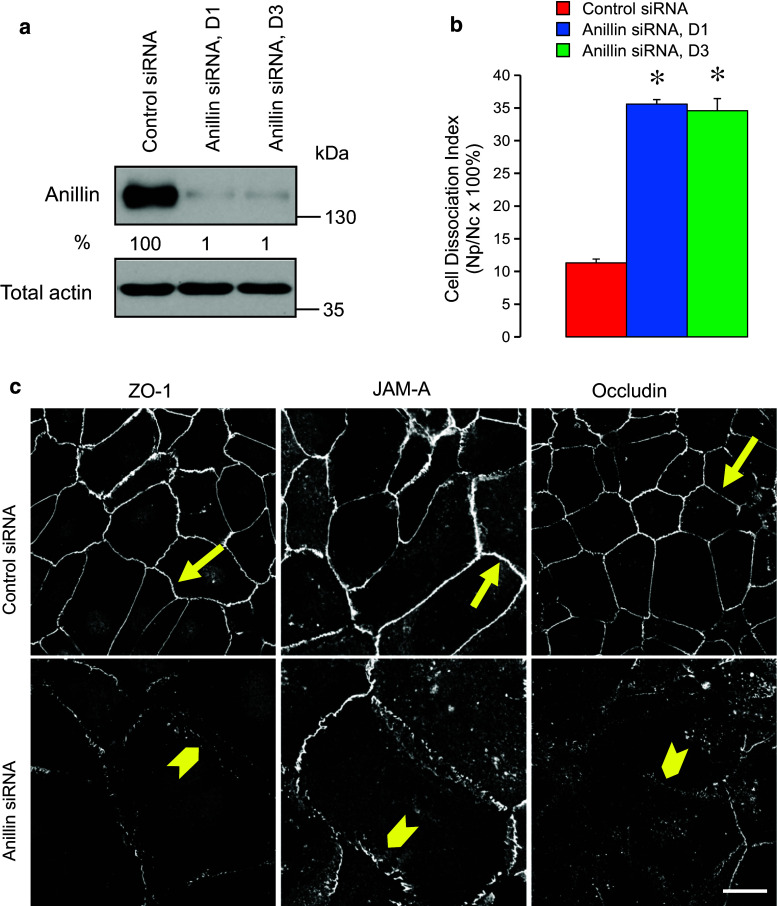
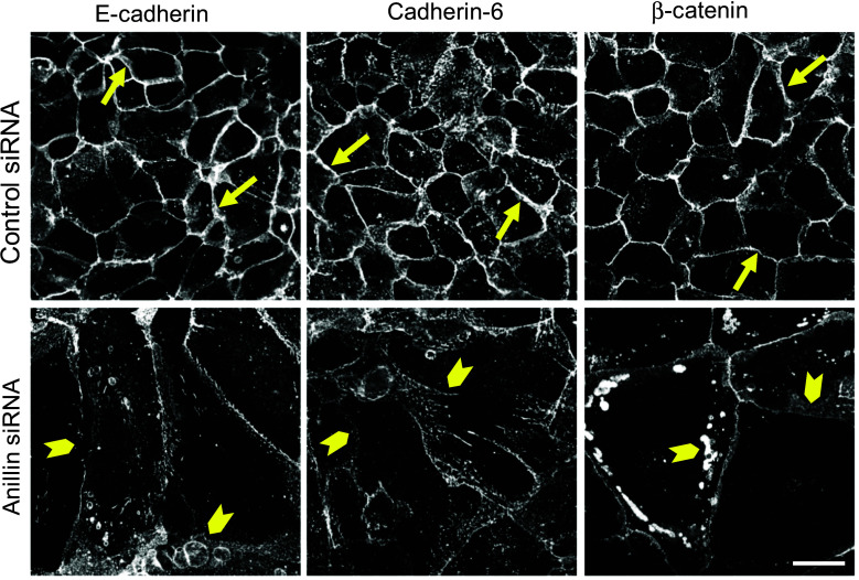
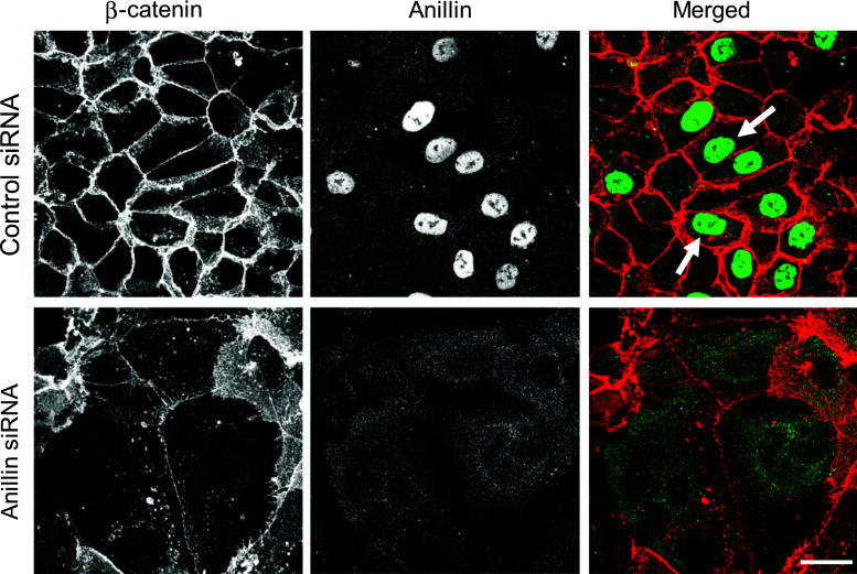
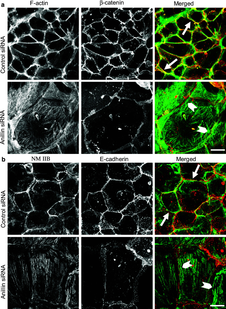
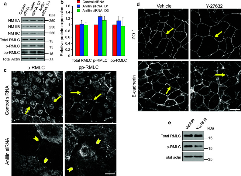
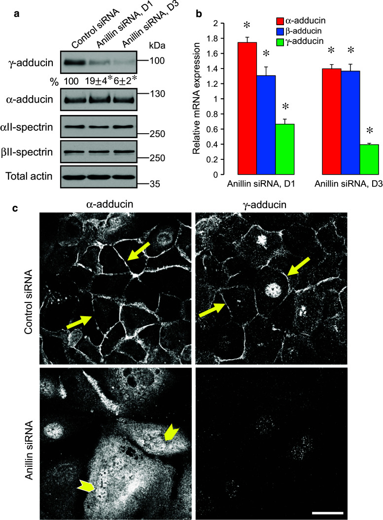
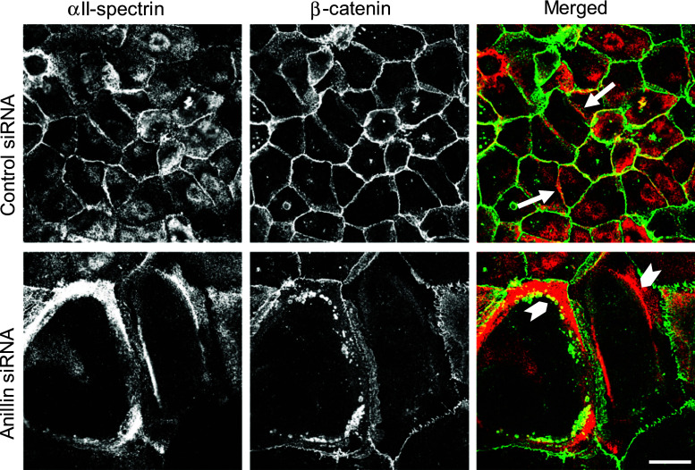
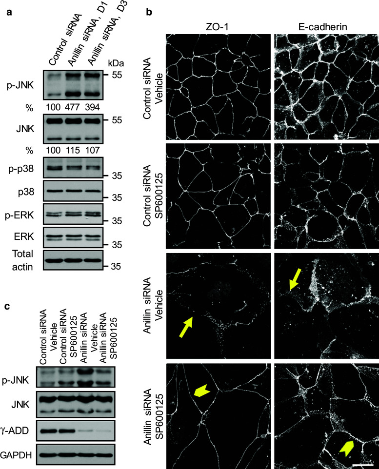
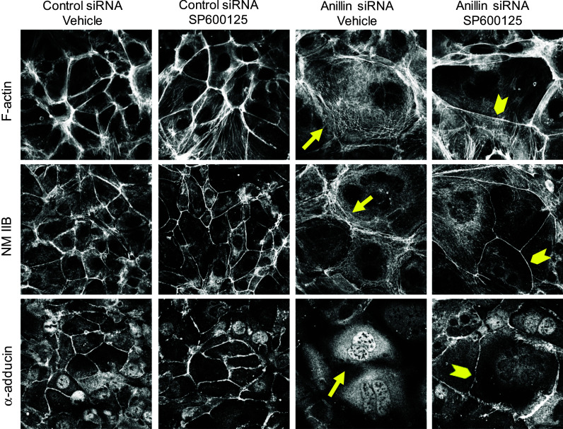
Tight junctions (TJ) and adherens junctions (AJ) are key morphological features of differentiated epithelial cells that regulate the integrity and permeability of tissue barriers. Structure and remodeling of epithelial junctions depends on their association with the underlying actomyosin cytoskeleton. Anillin is a unique scaffolding protein interacting with different cytoskeletal components, including actin filaments and myosin motors. Its role in the regulation of mammalian epithelial junctions remains unexplored. Downregulation of anillin expression in human prostate, colonic, and lung epithelial cells triggered AJ and TJ disassembly without altering the expression of junctional proteins. This junctional disassembly was accompanied by dramatic disorganization of the perijunctional actomyosin belt; while the general architecture of the actin cytoskeleton, and activation status of non-muscle myosin II, remained unchanged. Furthermore, loss of anillin disrupted the adducin-spectrin membrane skeleton at the areas of cell-cell contact, selectively decreased γ-adducin expression, and induced cytoplasmic aggregation of αII-spectrin. Anillin knockdown activated c-Jun N-terminal kinase (JNK), and JNK inhibition restored AJ and TJ integrity and cytoskeletal organization in anillin-depleted cells. These findings suggest a novel role for anillin in regulating intercellular adhesion in model human epithelia by mechanisms involving the suppression of JNK activity and controlling the assembly of the perijunctional cytoskeleton.


Figures

Fig. 1
Fig. 1
Anillin depletion weakens intercellular contacts and induces tight junction disassembly in prostate epithelial cells. DU145 cells were transfected with either control or two anillin-specific siRNA duplexes (D1 and D3) and examined on day 4 post-transfection. a Efficiency of anillin knockdown was determined by immunoblotting with quantitative densitometric analysis. b Avidity of intercellular contact was determined by cell dissociation assay. c Morphology of tight junctions was examined using immunofluorescent labeling of ZO-1, JAM-A, and occludin. Arrows indicate normal tight junction structure in control siRNA-transfected cells. Arrowheads highlight areas of tight junction disassembly in anillin-depleted cells. Data are presented as mean ± SE (n = 3); *p < 0.001. Scale bar 20 µm
Fig. 2
Fig. 2
Anillin depletion disrupts the integrity of epithelial adherens junctions. DU145 cells were transfected with either control or anillin-specific siRNAs and immunolabeled for adherens junction proteins E-cadherin, Cadherin 6, and β-catenin on day 4 post-transfection. Arrows indicate accumulation of all adherens junction proteins at the areas of cell–cell contact in control cells. Arrowheads highlight the disappearance of these proteins from intercellular junctions after anillin depletion. Scale bar 20 µm
Fig. 3
Fig. 3
Anillin does not localize at intercellular junctions in confluent epithelial cell monolayers. Control and anillin-depleted DU145 cell monolayers were subjected to dual immunolabeling for anillin (green) and β-catenin (red). Arrows indicate specific nuclear localization of anillin in control DU145 cells. This nuclear labeling disappears after siRNA-mediated anillin depletion. Scale bar 20 µm
Fig. 4
Fig. 4
Loss of anillin induces disorganization of the perijunctional actomyosin belt. DU145 cells transfected with either control or anillin-specific siRNAs were subjected to dual labeling for either a filamentous (F) actin (green) and β-catenin (red) or b NM IIB (green) and E-cadherin (red) on day 4 post-transfection. Arrows indicate a well-formed perijunctional actomyosin belt in control cells. Arrowheads highlight disorganized perijunctional actin filaments and basal NM IIB-enriched stress fibers in anillin-depleted cells. Scale bar 20 µm
Fig. 5
Fig. 5
Effects of anillin depletion on epithelial junctions are not mediated by altered NM II activity. a, b DU145 cells were transfected with either control or anillin-specific siRNAs (duplex 1 and 3) and were harvested for total cell lysates on day 4 post-transfection. Expression of different NM II heavy chain isoforms, total regulatory myosin light chain (RMLC), as well as monophosphorylated (p) and diphosphorylated (pp) RMLC, was determined by immunoblotting. c Control and anillin-depleted DU145 cells were immunolabeled for p-RMLC and pp-RMLC. Arrows point out localization of phosphorylated RMLC at epithelial junctions in control DU145 cells, whereas arrowheads highlight loss of such localization following anillin depletion. d, e Control DU145 cells were treated for 24 h with either vehicle, or ROCK inhibitor, Y-27632 (25 µM), and examined for AJ/TJ integrity and RMLC phosphorylation. Arrows indicate intact AJ and TJ in either vehicle or Y-27632-treated epithelial cells. Scale bar 20 µm
Fig. 6
Fig. 6
Anillin depletion alters expression and localization of adducins. Protein (a) and mRNA expression (b) of adducin and spectrin isoforms was examined in control and anillin-depleted DU145 cells on day 4 post-siRNA transfection. c Localization of α-adducin and γ-adducin was determined by immunofluorescence labeling. Arrows indicate accumulation of α-adducin and γ-adducin at intercellular junctions in control cells. Arrowheads highlight predominantly cytoplasmic labeling of α-adducin after anillin depletion. Data are presented as mean ± SE (n = 3); *p < 0.05. Scale bar 20 µm
Fig. 7
Fig. 7
Anillin depletion affects cellular distribution of α-spectrin. DU145 cells were transfected with either control or anillin-specific siRNAs and subjected to dual immunolabeling for αII-spectrin (red) and β-catenin (green). Arrows indicate localization of αII-spectrin at intercellular junctions in control cells. Arrowheads highlight accumulation of cytoplasmic αII-spectrin aggregates in anillin-depleted cells. Scale bar 20 µm
Fig. 8
Fig. 8
Activation of JNK mediates junctional disassembly in anillin-depleted epithelial cells. a Expression and phosphorylation of three major MAPKs was determined in control and anillin-depleted DU145 cells via immunoblotting. b, c Control and anillin-depleted DU145 cells were treated for 24 h with either vehicle or JNK inhibitor, SP600125 (25 µM). The effect of JNK inhibition on AJ and TJ integrity was determined by immunolabeling of E-cadherin and ZO-1. JNK activation and expression of γ-adducin was examined by immunoblotting. Arrows indicate AJ/TJ disassembly in vehicle-treated anillin-depleted cells. Arrowheads highlight restoration of junctional integrity of anillin-depleted cells by JNK inhibition. Scale bar 20 µm
Fig. 9
Fig. 9
JNK inhibition restores the perijunctional actomyosin belt and α-adducin localization in anillin-depleted cells. Control and anillin-depleted DU145 cells were treated for 24 h with either vehicle or JNK inhibitor, SP600125 (25 µM). The effect of JNK inhibition on the perijunctional actomyosin cytoskeleton and the membrane skeleton was determined by fluorescence labeling. Arrows indicate disruption of F-actin, NM IIB, and α-adducin localization at intercellular contacts of vehicle-treated anillin-depleted cells. Arrowheads highlight restoration of the perijunctional actomyosin belt and junction-associated α-adducin fraction in anillin-depleted cells following JNK inhibition. Scale bar 20 µm

Similar articles

Cited by

References

    1. Anderson JM, Van Itallie CM. Physiology and function of the tight junction. Cold Spring Harb Perspect Biol. 2009;1:a002584. doi: 10.1101/cshperspect.a002584. - DOI - PMC - PubMed
    1. Cheng CY, Mruk DD. The blood-testis barrier and its implications for male contraception. Pharmacol Rev. 2012;64:16–64. doi: 10.1124/pr.110.002790. - DOI - PMC - PubMed
    1. Turner JR. Intestinal mucosal barrier function in health and disease. Nat Rev Immunol. 2009;9:799–809. doi: 10.1038/nri2653. - DOI - PubMed
    1. Green KJ, Getsios S, Troyanovsky S, Godsel L. Intercellular junction assembly, dynamics, and homeostasis. Cold Spring Harb Perspect Biol. 2010;2:a000125. doi: 10.1101/cshperspect.a000125. - DOI - PMC - PubMed
    1. Tsukita S, Furuse M, Itoh M. Multifunctional strands in tight junctions. Nat Rev Mol Cell Biol. 2001;2:285–293. doi: 10.1038/35067088. - DOI - PubMed

Publication types

MeSH terms