. 2015 Mar 24;108(6):1484-1494.
doi: 10.1016/j.bpj.2015.02.012.

LKB1/Mo25/STRAD uniquely impacts sarcomeric contractile function and posttranslational modification

Affiliations

LKB1/Mo25/STRAD uniquely impacts sarcomeric contractile function and posttranslational modification

Samantha M Behunin et al. Biophys J. .

Abstract

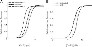
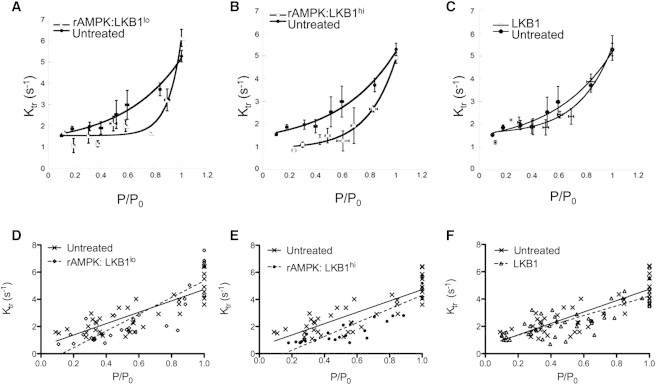
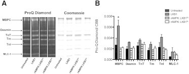
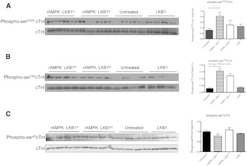
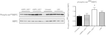
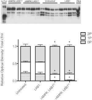
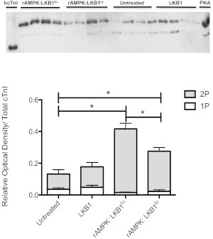
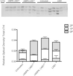
The myocardium undergoes extensive metabolic and energetic remodeling during the progression of cardiac disease. Central to remodeling are changes in the adenine nucleotide pool. Fluctuations in these pools can activate AMP-activated protein kinase (AMPK), the central regulator of cellular energetics. Binding of AMP to AMPK not only allosterically activates AMPK but also promotes phosphorylation of AMPK by an upstream kinase complex, LKB1/Mo25/STRAD (liver kinase B 1, mouse protein 25, STE-related adaptor protein). AMPK phosphorylation by the LKB1 complex results in a substantial increase in AMPK activity. Molecular targeting by the LKB1 complex depends on subcellular localization and transcriptional expression. Yet, little is known about the ability of the LKB1 complex to modulate targeting of AMPK after activation. Accordingly, we hypothesized that differing stoichiometric ratios of LKB1 activator complex to AMPK would uniquely impact myofilament function. Demembranated rat cardiac trabeculae were incubated with varying ratios of the LKB1 complex to AMPK or the LKB1 complex alone. After incubation, we measured the Ca(2+) sensitivity of tension, rate constant for tension redevelopment, maximum tension generation, length-dependent activation, cooperativity, and sarcomeric protein phosphorylation status. We found that the Ca(2+) sensitivity of tension and cross-bridge dynamics were dependent on the LKB1 complex/AMPK ratio. We also found that the LKB1 complex desensitizes and suppresses myofilament function independently of AMPK. A phospho-proteomic analysis of myofilament proteins revealed site-specific changes in cardiac Troponin I (cTnI) phosphorylation, as well as a unique distribution of cTnI phosphospecies that were dependent on the LKB1 complex/ AMPK ratio. Fibers treated with the LKB1 complex alone did not alter cTnI phosphorylation or phosphospecies distribution. However, LKB1 complex treatment independent of AMPK increased phosphorylation of myosin-binding protein C. Therefore, we conclude that the LKB1/AMPK signaling axis is able to alter muscle function through multiple mechanisms.


Figures

Figure 1
Figure 1
Ca2+ sensitivity of tension development in untreated and treated demembranated cardiac trabeculae. (A) Ca2+ sensitivity of tension in trabeculae treated with low levels of LKB1 complex activation of AMPK (open diamonds, rAMPK/LKB1lo) and high levels of LKB1 complex activation (open squares, rAMPK/LKB1hi) compared with untreated fibers (solid circles, untreated). (B) Ca2+ sensitivity of tension in trabeculae treated with only the LKB1 complex (open circles) compared with untreated fibers (solid circles). SL was set to 2.2 μm and all data were normalized to saturating Ca2+ (maximal) tension.
Figure 2
Figure 2
Rate constant for tension redevelopment. ktr is plotted as a function of normalized tension for fibers treated with low LKB1 activation of AMPK (rAMPK/LKB1lo), high LKB1 activation of AMPK (rAMPK/LKB1hi), and LKB1 complex alone (LKB1) plotted relative to untreated fibers. (A–C) Data were binned and fitted to a curvilinear fit. There is a significant increase in the slope of rAMPK/LKB1lo-treated fibers compared with untreated fibers (p < 0.05). rAMPK/LKB1hi-treated fibers tend to have an increased slope compared with untreated fibers. There is no difference between curvilinear slopes between LKB1-complex-treated fibers and untreated fibers. (D–F) Raw data were fitted with a linear regression, and multiple linear regression analysis was used to measure differences between slopes. There is a significant increase in the slope in fibers treated with rAMPK/LKB1lo (p < 0.05). There is a tendency for an increased slope with rAMPK/LKB1hi-treated fibers, with a significantly different intercept (p < 0.05) compared with untreated fibers. There is no difference in linear slopes between LKB1-complex-treated and untreated fibers.
Figure 3
Figure 3
ProQ Diamond phosphoprotein stain after treatment with and without various activated AMPKs and the LKB1 complex. (A) Representative lanes (n = 4 per treatment group) from the same gel stained first for phosphorylation (ProQ Diamond) and then for total protein content (Coommassie Brilliant Blue). Protein identification as shown on the left was based on the molecular weights of known myofibrillar proteins. (B) The relative optical density of the phosphoprotein signal was normalized for loading by dividing the ProQ Diamond signal by the corresponding Coomassie Brilliant Blue signal per protein. There is a significant increase in phosphorylation of MBPC in skinned trabeculae treated with only exogenous LKB1 complex compared with all other groups (p < 0.05; n = 4 per group). In all other proteins, there is no significant change in global phosphorylation status.
Figure 4
Figure 4
Western blot analysis of phosphorylated cTnI. The Western blot depicts phospho-cTnI in myofibrils treated with various stoichiometric LKB1 activations of AMPK and the LKB1 complex, with the corresponding normalized optical density plotted on the right. (A) Phospho-ser23/24 cTnI was measured in treated and untreated myofibrils. Fibers treated with a high level of LKB1 activation of AMPK (rAMPK/LKB1hi) had a significant increase in phosphorylated ser23/24 content relative to untreated myofibrils (p < 0.05; n = 4 for all groups). (B) Phospho-ser150 cTnI was measured in treated and untreated myofibrils. There was a significant increase in phosphorylated ser150 in rAMPK/LKB1hi- and rAMPK/LKB1lo-treated fibers (p < 0.05; n = 3–4 per group). (C) Phospho-ser43cTnI was measured in treated and untreated myofibrils. There was no change in phospho-ser43cTnI in any treatment group (p > 0.05).
Figure 5
Figure 5
Western blot analysis of phosphorylated MBPC. The Western blot depicts phospho-ser282MPBC in myofibrils treated with activated AMPK and the LKB1 complex. The phosphorylated signal was normalized to total MBPC, and the corresponding normalized optical density is plotted on the right. Fibers treated with a low level of LKB1 activation of AMPK (rAMPK/LKB1lo) had a significant increase in phosphorylated ser282 content relative to untreated myofibrils (p < 0.05; n = 4 for all groups).
Figure 6
Figure 6
Phosphate-affinity SDS-PAGE (Phos-tag) of the total cTnI phosphospecies distribution. cTnI was separated into unphosphorylated [0P], monophosphorylated [1P], and bisphosphorylated [2P] phosphospecies by SDS-PAGE-Phos-tag followed by Western blot analysis with a total cTnI antibody. Top panel: representative SDS-PAGE-Phos-tag followed by Western blot, illustrating three bands corresponding to 0P, 1P, and 2P. Human recombinant cTnI (hcTnI) and PKA-treated (+PKA) cardiomyocytes were used to confirm each phosphospecies. Bottom panel: stacked bar graph indicating the relative amounts of each phosphospecies per experimental group. There is a significant shift in fibers treated with both high and low LKB1 activation of AMPK (rAMPK/LKB1hi and rAMPK:LKB1lo) away from the 0P state (p < 0.05). There is no change in total cTnI phosphospecies distribution in the LKB1-complex-treated group.
Figure 7
Figure 7
Phosphate-affinity SDS-PAGE (Phos-tag) of the phospho-ser23/24 cTnI phosphospecies distribution. cTnI was separated into unphosphorylated [0P], monophosphorylated [1P], and bisphosphorylated [2P] phosphospecies by SDS-PAGE-Phos-tag followed by Western blot analysis with a phospho-ser23/24 cTnI antibody. Top panel: representative SDS-PAGE-Phos-tag followed by Western blot, illustrating two distinct bands corresponding to 1P and 2P. Human recombinant cTnI (hcTnI) and PKA-treated (+PKA) cardiomyocytes were used to confirm each phosphospecies. Bottom panel: stacked bar graph indicating the relative amounts of each phosphospecies per experimental group. There is a significant increase in 2P phospho-ser23/24cTnI in rAMPK/LKB1hi- and rAMPK/LKB1lo-treated fibers (p < 0.05). In addition, rAMPK/LKB1hi-treated fibers contain more 2P phospho-ser23/24cTnI than rAMPK/LKB1lo-treated fibers. There was no change in the group treated only with LKB1 complex.
Figure 8
Figure 8
Phosphate-affinity SDS-PAGE (Phos-tag) of the phospho-ser150 cTnI phosphospecies distribution. cTnI was separated into unphosphorylated [0P], monophosphorylated [1P], and bisphosphorylated [2P] phosphospecies by SDS-PAGE-Phos-tag followed by Western blot analysis with a phospho-ser150 cTnI antibody. Top: representative SDS-PAGE-Phos-tag followed by Western blot illustrating two or three distinct bands corresponding to 0P, 1P, and 2P. hcTnI was used as the unphosphorylated cTnI standard. Bottom: stacked bar graph indicating the relative amounts of each phosphospecies per experimental group. There was no change in the distribution of phospho-ser150 cTnI phosphospecies among groups.

References

    1. Doenst T., Nguyen T.D., Abel E.D. Cardiac metabolism in heart failure: implications beyond ATP production. Circ. Res. 2013;113:709–724. - PMC - PubMed
    1. Ingwall J.S. Energy metabolism in heart failure and remodelling. Cardiovasc. Res. 2009;81:412–419. - PMC - PubMed
    1. Nascimben L., Ingwall J.S., Allen P.D. Creatine kinase system in failing and nonfailing human myocardium. Circulation. 1996;94:1894–1901. - PubMed
    1. Aksentijević D., Lygate C.A., Neubauer S. High-energy phosphotransfer in the failing mouse heart: role of adenylate kinase and glycolytic enzymes. Eur. J. Heart Fail. 2010;12:1282–1289. - PMC - PubMed
    1. Dzeja P., Terzic A. Adenylate kinase and AMP signaling networks: metabolic monitoring, signal communication and body energy sensing. Int. J. Mol. Sci. 2009;10:1729–1772. - PMC - PubMed

Publication types

MeSH terms