. 2015 Mar 24;108(6):1503-1515.
doi: 10.1016/j.bpj.2015.01.039.

Probing the structural and dynamical effects of the charged residues of the TZF domain of TIS11d

Affiliations

Probing the structural and dynamical effects of the charged residues of the TZF domain of TIS11d

Brittany R Morgan et al. Biophys J. .

Abstract

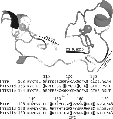
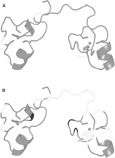
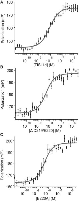
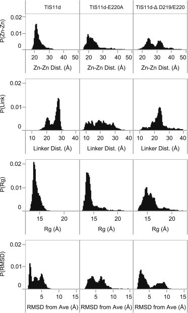
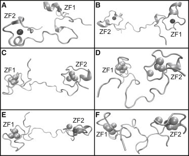
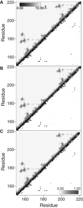
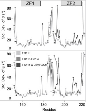
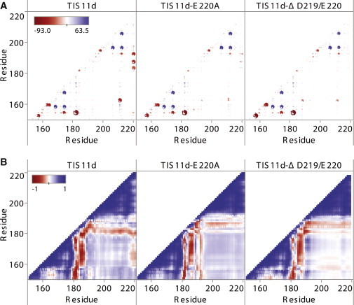
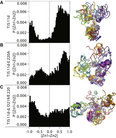
A member of the TTP family of proteins, TIS11d binds RNA with high specificity using a pair of CCCH-type tandem zinc fingers separated by a 18 residue long linker. Our previous work showed that the formation of hydrogen bonds between the C-terminal residue E220 and the residues of the linker region stabilized a compact structure of TIS11d in the absence of RNA. To investigate the role of the C-terminal residues in the structure of unbound TIS11d, the E220A mutant and the truncation mutant lacking the last two residues (D219/E220) were studied using molecular dynamics, NMR spectroscopy, and biochemical methods. This study confirmed the importance of the charged residues D219 and E220 in maintaining structural stability in unbound TIS11d and elucidated the underlying physical mechanisms. We observed a greater structural heterogeneity for the residues of the linker in the molecular dynamics trajectories of both mutant proteins relative to the wild-type. This heterogeneity was more pronounced in the D219/E220 deletion mutant than in the E220A mutant, indicating that a greater reduction of the charge of the C-terminus results in greater flexibility. In agreement with the increased flexibility and the reduced number of negatively charged residues of the D219/E220 deletion mutant, we measured more unfavorable entropic and a more favorable enthalpic contribution to the free energy of RNA binding in the mutant than in the wild-type protein. The relative orientation of the zinc fingers was stabilized by the electrostatic interaction between E220 and positively charged residues of the linker in TIS11d. In the E220A mutant, the relative orientation of the zinc fingers was less constrained, whereas in the D219/E220 deletion mutant, little orientational preference was observed. We posit that favorable electrostatic interactions provide a mechanism to promote preferential orientation of separate domains without imposing structural rigidity.


Figures

Figure 1
Figure 1
Structure and sequence alignment of the RBD of TIS11d. Top: representation of TIS11d (PDB access code 1RGO, RNA not shown) colored by charge (black: negative residues, white: positive residues). Gray shows backbone (tube), Zn2+ ions (spheres). Bottom: sequence alignment of the RBD of TIS11 family members with total charge.
Figure 2
Figure 2
Comparison of 15N-1H HSQC spectra. Dots on top of the spectrum indicate whether a crosspeak was observed for each residue. (A) TIS11d (gray) and TIS11d-E220A (black), (B) TIS11d (gray) and TIS11d-Δ D219/E220 (black).
Figure 3
Figure 3
Crosspeaks with lost or reduced signal in the 15N-1H HSQC spectra of TIS11-E220A and TIS11d-Δ D219/E220 relative to WT TIS11d mapped on the initial NMR structure. Gray: residues with no significant change, black: residues with reduced signal, white: residues with no signal above the noise. (A) TIS11d-E220A and (B) TIS11d-Δ D219/E220.
Figure 4
Figure 4
Thermodynamics of protein/RNA binding. Fluorescence polarization data used to calculate Kd,app of RNA-binding for (A) TIS11d, (B) TIS11d-Δ D219/E220, (C) TIS11d-E220A.
Figure 5
Figure 5
Normalized histograms of combined trajectories of structural metrics for each system (as labeled). From top to bottom: distance between zinc ions, linker end-to-end distance, radius of gyration, and RMSD from average structure.
Figure 6
Figure 6
Example structures of TIS11d-E220A (A and B) and TIS11d-Δ D219/E220 (CF). Black spheres: zinc ions, gray spheres: sulfur atoms of coordinating cysteine residues (large spheres) and nitrogen residues (small spheres), gray ribbon: backbone. TIS11d-E220A: (A) before extension, (B) during extension. TIS11d-Δ D219/E220: (C) extension with zinc ion of ZF2 still coordinated to Cys-212, (D) after extension with loss of Cys-212 coordination, (E) extended structure with loss of Cys-212 coordination, (F) more compact structure with crossed linker structure.
Figure 7
Figure 7
Above the diagonal: backbone distance maps by system averaged over all trajectories. The mean distance between Cα atoms of each residue pair is shown from dark (0 Å) to light (10 Å cutoff). Below the diagonal: hydrogen bond probability maps by system averaged over all trajectories. (A) TIS11d, (B) TIS11d-E220A, (C) TIS11d-Δ D219/E220.
Figure 8
Figure 8
SD of backbone dihedral angles over all trajectories of TIS11d (light gray), TIS11d-E220A (medium gray), and TIS11d-Δ D219/E220 (dark gray).
Figure 9
Figure 9
(A) Interaction energy between residue pairs by system (as labeled) averaged over all trajectories. Red: negative (favorable) interaction energies, blue: positive (unfavorable) interaction energies. Size of circles is a function of the interaction strength to highlight strong interactions, the larger the circle the stronger the interaction. (B) Cross correlation between residues when aligned on residues 179–190 by system (as labeled) averaged over all trajectories. Blue is positively correlated, red is anticorrelated.
Figure 10
Figure 10
Characterization of zinc finger relative orientation for each system (as labeled). Left: normalized histograms of zinc finger domain relative orientation. Right: representative structures colored by time from trajectory 1 for each system aligned on residues 179–190. Red-yellow structures are early time points in the trajectories, orange-green structures are mid trajectory, and blue-purple structures are toward the end of the trajectories.

Similar articles

Cited by

References

    1. Carballo E., Lai W.S., Blackshear P.J. Feedback inhibition of macrophage tumor necrosis factor-alpha production by tristetraprolin. Science. 1998;281:1001–1005. - PubMed
    1. Chen C.Y., Gherzi R., Karin M. AU binding proteins recruit the exosome to degrade ARE-containing mRNAs. Cell. 2001;107:451–464. - PubMed
    1. Lai W.S., Carballo E., Blackshear P.J. Evidence that tristetraprolin binds to AU-rich elements and promotes the deadenylation and destabilization of tumor necrosis factor alpha mRNA. Mol. Cell. Biol. 1999;19:4311–4323. - PMC - PubMed
    1. Blackshear P.J. Tristetraprolin and other CCCH tandem zinc-finger proteins in the regulation of mRNA turnover. Biochem. Soc. Trans. 2002;30:945–952. - PubMed
    1. Brewer B.Y., Malicka J., Wilson G.M. RNA sequence elements required for high affinity binding by the zinc finger domain of tristetraprolin: conformational changes coupled to the bipartite nature of Au-rich MRNA-destabilizing motifs. J. Biol. Chem. 2004;279:27870–27877. - PubMed

Publication types

Substances

LinkOut - more resources