. 2015 Feb 10:12:15.
doi: 10.1186/s12977-015-0135-y.

The HIV-1 antisense protein (ASP) induces CD8 T cell responses during chronic infection

The HIV-1 antisense protein (ASP) induces CD8 T cell responses during chronic infection

Anne Bet et al. Retrovirology. .

Abstract

Background: CD8+ T cells recognize HIV-1 epitopes translated from a gene's primary reading frame (F1) and any one of its five alternative reading frames (ARFs) in the forward (F2, F3) or reverse (R1-3) directions. The 3' end of HIV-1's proviral coding strand contains a conserved sequence that is directly overlapping but antiparallel to the env gene (ARF R2) and encodes for a putative antisense HIV-1 protein called ASP. ASP expression has been demonstrated in vitro using HIV-transfected cell lines or infected cells. Although antibodies to ASP were previously detected in patient sera, T cell recognition of ASP-derived epitopes has not been evaluated. We therefore investigated the ex vivo and in vitro induction of ASP-specific T cell responses as a measure of immune recognition and protein expression during HIV-1 infection.

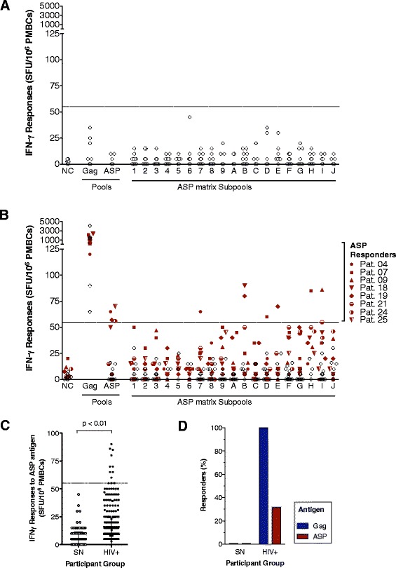
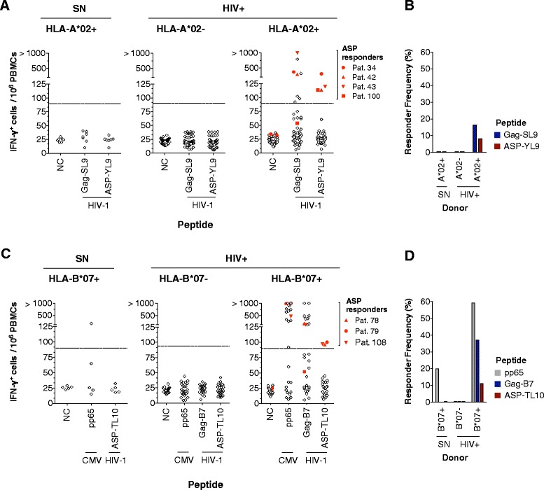
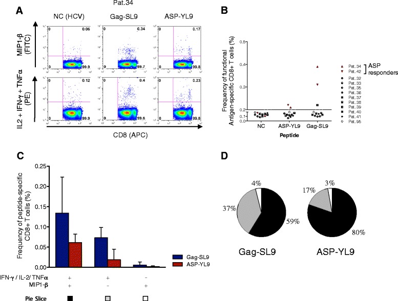
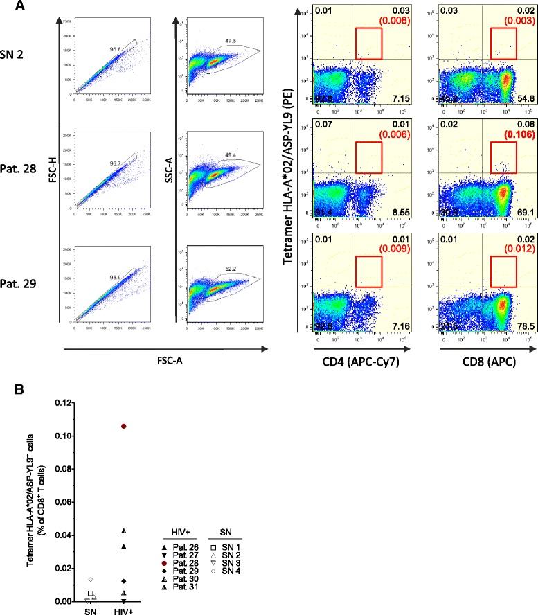
Results: A panel of overlapping peptides was initially designed from the full-length ASP sequence to perform a global assessment of T cell responses. Recognition of ASP-derived antigens was evaluated in an IFN-γELISpot assay using PBMCs from HIV-1 seropositive and seronegative individuals. Eight of 25 patients had positive responses to ASP antigens and none of the seronegative donors responded. As a complimentary approach, a second set of antigens was designed using HLA-I binding motifs and affinities. Two ASP-derived peptides with high predicted binding affinities for HLA-A*02 (ASP-YL9) and HLA-B*07 (ASP-TL10) were tested using PBMCs from HIV-1 seropositive and seronegative individuals who expressed the matching HLA-I-restricting allele. We found that HLA-I-restricted ASP peptides were only recognized by CD8+ T cells from patients with the relevant HLA-I and did not induce responses in any of the seronegative donors or patients who do not express the restrictive HLA alleles. Further, ASP-YL9-specific CD8+ T cells had functional profiles that were similar to a previously described HLA-A*02-restricted epitope (Gag-SL9). Specific recognition of ASP-YL9 by CD8+ T cells was also demonstrated by tetramer staining using cells from an HLA-A*02 HIV-infected patient.

Conclusion: Our results provide the first description of CD8+ T cell-mediated immune responses to ASP in HIV-1-infected patients, demonstrating that ASP is expressed during infection. Our identification of epitopes within ASP has implications for designing HIV vaccines.


Figures

Figure 1
Figure 1
HIV-1 ASP peptide pools activate IFN-γ T cell responses in a cohort of HIV+ individuals. The magnitude of IFN-γ ELISpot responses (SFU/106 PBMCs) to the Gag or ASP pools and subpools (1 to 9, and A to J) are shown for (A) seronegative donors and (B) HIV-1+ patients. Each dot represents one donor and mean IFN-γ responses of triplicate or quadruplicate are indicated. The dashed line marks the standardized threshold (55 SFU/106 PBMCs) for positivity. (C) PBMCs were assessed for IFN-γ production using ASP pools and 19 ASP subpools. Mann–Whitney U tests were performed to determine significant differences between the median responses to peptide pools (p < 0.01). (D) The frequency of responders is illustrated as a proportion of seronegative (SN) or seropositive (HIV+) individuals with positive responses to the Gag pool (blue column) and either to the ASP pool or any ASP subpool (red column).
Figure 2
Figure 2
HLA-I-restricted ASP epitopes are recognized by CD8+ T cells during HIV-1 infection. (A) PBMCs isolated from HIV-1 seropositive patients and seronegative donors were stimulated ex vivo with ASP-YL9 in an IFN-γ ELISpot assay. An HCV irrelevant and HIV Gag-derived HLA-A*02-restricted (Gag-SL9) peptide was used as negative and positive controls, respectively. Dotted line indicates a mean threshold of peptide responders (4 times background, 90 SFU/106 PBMCs). Four HLA-A*02+ HIV+ patients had responses to ASP-YL9 (n = 49, right panel), whereas no IFN-γ secretion was detected in HLA-A*02- HIV+ patients (n = 30, middle panel) or HLA-A*02+ seronegative donors (SN, n = 7, left panel). (B) Frequencies of ASP-YL9-, or Gag-SL9- responders according to each group are shown. (C) As in (B) with the ASP-TL10 peptide and a CMV-derived (pp65) peptide. Three HLA-B*07+ HIV+ donors were responding to ASP-TL10 (n = 27, right panel), whereas no responses were detected in HLA-B*07- seropositive donors (n = 27, middle panel) and HLA-B*07+ SN (n = 5, left panel). (D) Frequencies of donors responding to ASP-TL10, HIV Gag (Gag-B7) or CMV (pp65) are indicated according to each group.
Figure 3
Figure 3
The HIV-1 ASP-YL9 peptide activates CD8 + T cell responses. Intracellular cytokine staining (ICS) was used to analyze CD8+ T cell responses to ASP-YL9 in 11 HLA-A*02+ patients (Pats.32 to 42) and one HLA-A*02- patient (Pat.95). (A) Flow cytometry analysis from Pat.34, illustrating MIP1-β versus IL-2/TNFα/IFN-γ production by CD8+ cells after HCV- (negative control, NC), HIV ASP-YL9 or HIV Gag-SL9 stimulation. (B) Frequencies of CD8+ cells producing at least one of the analyzed cytokine/chemokine. Pats. 34 and 42 were activated upon ASP-YL9 stimulation. (C) For these two patients, percentages of Gag-SL9 (blue) or ASP-YL9 (red) stimulated CD8+ cells, producing either only MIP1-β (white), only IL-2/TNFα/IFN-γ (grey), or both chemokines/cytokines panels (black) were analyzed. NC background was subtracted and average percentages (±SD) from Pats.34 and 42 analysis are presented. (D) Pies indicate relative proportions of Gag-SL9 or ASP-YL9 activated CD8+ T cells producing each panel of cytokines/chemokines (presented in C).
Figure 4
Figure 4
Detection by tetramer of HLA-A*02-restricted, ASP-YL9-specific CD8+ T cells in HIV+ patients. Following in vitro restimulation of PBMCs with ASP-YL9 peptide, cell lines from 6 HLA-A*02+ HIV+ patients (Pats. 26 to 31) and 4 HLA-A*02+ seronegative donors (SN 1 to 4) were stained for CD4 and CD8 in combination with the HLA-A*02/ASP-YL9 tetramer. (A) Gating strategies and illustration of tetramer staining for SN2, Pat.28 and Pat.29. In Pat.28, 0.06% tetramers + cells were detected corresponding to 0.11% of CD8+ T cells. No tetramer + cells were detected among CD4+ cells. In Pat.29, no significant tetramer+ population was detected (Fisher-exact test). (B) Percentages of HLA-A*02/ASP-YL9 tetramer+ cells within CD8+ T cells of HIV+ patients and seronegative donors.

References

    1. Schmitz JE, Kuroda MJ, Santra S, Sasseville VG, Simon MA, Lifton MA, et al. Control of viremia in simian immunodeficiency virus infection by CD8+ lymphocytes. Science. 1999;283:857–60. doi: 10.1126/science.283.5403.857. - DOI - PubMed
    1. Jin X, Bauer DE, Tuttleton SE, Lewin S, Gettie A, Blanchard J, et al. Dramatic rise in plasma viremia after CD8 (+) T cell depletion in simian immunodeficiency virus-infected macaques. J Exp Med. 1999;189:991–8. doi: 10.1084/jem.189.6.991. - DOI - PMC - PubMed
    1. Borrow P, Lewicki H, Hahn BH, Shaw GM, Oldstone MB. Virus-specific CD8+ cytotoxic T-lymphocyte activity associated with control of viremia in primary human immunodeficiency virus type 1 infection. J Virol. 1994;68:6103–10. - PMC - PubMed
    1. Wang YE, Li B, Carlson JM, Streeck H, Gladden AD, Goodman R, et al. Protective HLA class I alleles that restrict acute-phase CD8+ T-cell responses are associated with viral escape mutations located in highly conserved regions of human immunodeficiency virus type 1. J Virol. 2009;83:1845–55. doi: 10.1128/JVI.01061-08. - DOI - PMC - PubMed
    1. Wilson NA, Keele BF, Reed JS, Piaskowski SM, MacNair CE, Bett AJ, et al. Vaccine-induced cellular responses control simian immunodeficiency virus replication after heterologous challenge. J Virol. 2009;83:6508–21. doi: 10.1128/JVI.00272-09. - DOI - PMC - PubMed

Publication types