. 2015 May 8;290(19):12027-39.
doi: 10.1074/jbc.M114.608570. Epub 2015 Mar 25.

The Non-catalytic B Subunit of Coagulation Factor XIII Accelerates Fibrin Cross-linking

Affiliations

The Non-catalytic B Subunit of Coagulation Factor XIII Accelerates Fibrin Cross-linking

Masayoshi Souri et al. J Biol Chem. .

Abstract

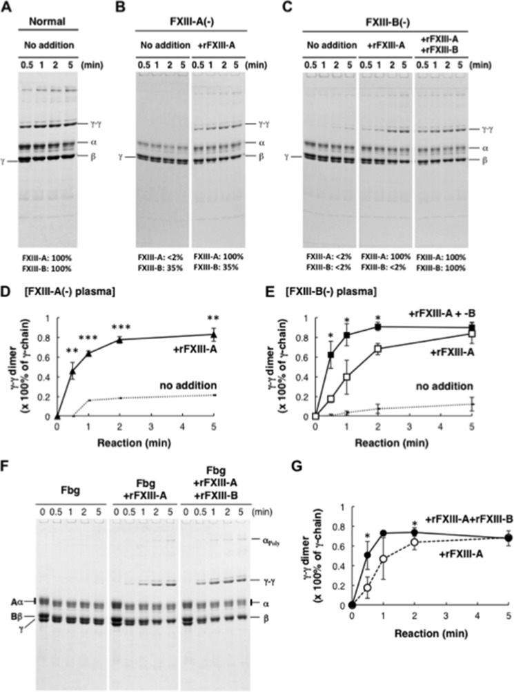
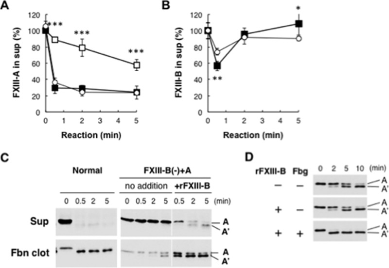
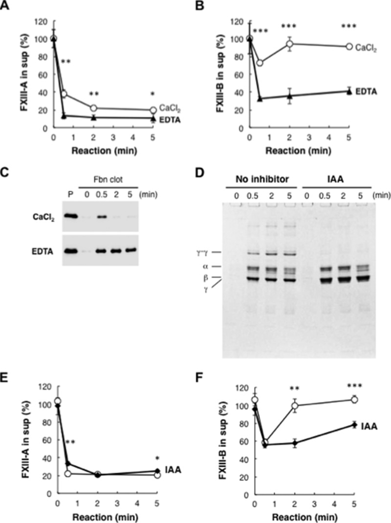
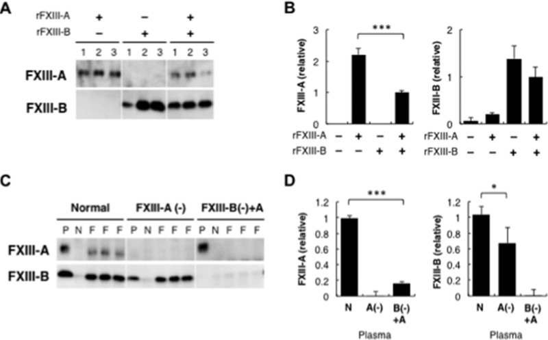
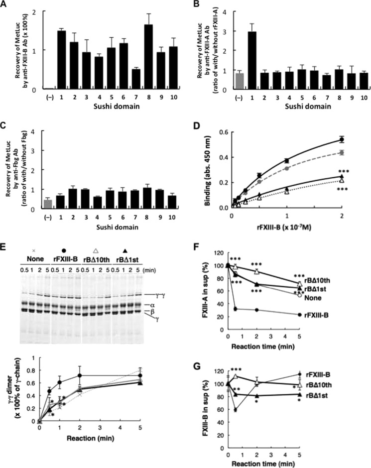
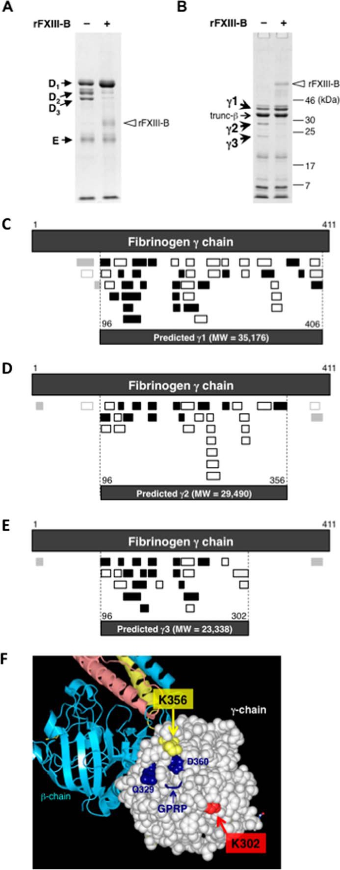
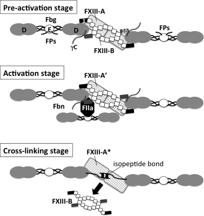
Covalent cross-linking of fibrin chains is required for stable blood clot formation, which is catalyzed by coagulation factor XIII (FXIII), a proenzyme of plasma transglutaminase consisting of catalytic A (FXIII-A) and non-catalytic B subunits (FXIII-B). Herein, we demonstrate that FXIII-B accelerates fibrin cross-linking. Depletion of FXIII-B from normal plasma supplemented with a physiological level of recombinant FXIII-A resulted in delayed fibrin cross-linking, reduced incorporation of FXIII-A into fibrin clots, and impaired activation peptide cleavage by thrombin; the addition of recombinant FXIII-B restored normal fibrin cross-linking, FXIII-A incorporation into fibrin clots, and activation peptide cleavage by thrombin. Immunoprecipitation with an anti-fibrinogen antibody revealed an interaction between the FXIII heterotetramer and fibrinogen mediated by FXIII-B and not FXIII-A. FXIII-B probably binds the γ-chain of fibrinogen with its D-domain, which is near the fibrin polymerization pockets, and dissociates from fibrin during or after cross-linking between γ-chains. Thus, FXIII-B plays important roles in the formation of a ternary complex between proenzyme FXIII, prosubstrate fibrinogen, and activator thrombin. Accordingly, congenital or acquired FXIII-B deficiency may result in increased bleeding tendency through impaired fibrin stabilization due to decreased FXIII-A activation by thrombin and secondary FXIII-A deficiency arising from enhanced circulatory clearance.

Keywords: Bleeding Disease; Blood; Coagulation Factor; Fibrin; Protein Cross-linking; Transglutaminase.


Figures

FIGURE 1.
FIGURE 1.
Cross-linking of Fbn in the absence of FXIII-B. A–E, FXIII-A or FXIII-B was removed from normal human plasma using anti-FXIII-A or -B antibody-Sepharose. Cross-linking reactions were performed in normal (A), FXIII-A-depleted (B), and FXIII-B-depleted plasma (C) with or without the addition of rFXIII-A and rFXIII-B. Monomer and dimer of γ-chain in FXIII-A-depleted (D) and FXIII-B-depleted plasma (E) were quantified by a densitometer, and γ-γ dimer formation was calculated as dimer/(monomer + dimer). The mean of three independent experiments was plotted. Dotted line, no addition; filled triangle, FXIII-A-depleted plasma with rFXIII-A; open square, FXIII-B-depleted plasma with rFXIII-A; filled square, FXIII-B-depleted plasma with rFXIII-A and rFXIII-B, error bars, S.D. F and G, purified human Fbg was reacted with thrombin in the presence of rFXIII-A without or with rFXIII-B. Densitometric analyses of γ-γ dimer formation in three independent experiments were performed. Open circle, Fbg with rFXIII-A; filled circle, Fbg with rFXIII-A and rFXIII-B. *, p < 0.05; **, p < 0.01; ***, p < 0.001.
FIGURE 2.
FIGURE 2.
FXIII in Fbn clots from FXIII-B(−)+A plasma. FXIII-A (A) and -B (B) in the supernatant separated from the Fbn clot after the cross-linking reaction were quantified by ELISA, and their amounts are given as a percentage of unreacted plasma. The mean ± S.D. (error bars) of 3–5 independent experiments was plotted. Open circle, normal plasma; open square, FXIII-B(−)+A plasma (FXIII-B-depleted plasma supplemented with rFXIII-A); filled square, FXIII-B(−)+A plasma with rFXIII-B. C, cleavage of the activation peptide of FXIII-A during Fbn cross-linking in FXIII-B(-)+A plasma. FXIII-A in the supernatant and Fbn clot was visualized by Western blotting using an anti-FXIII-A antibody. D, effect of FXIII-B on cleavage of the activation peptide of FXIII-A in a reaction with purified Fbg. Purified human Fbg was reacted with thrombin in the presence of rFXIII-A without or with rFXIII-B, and then samples were analyzed by Western blotting using an anti-FXIII-A antibody. *, p < 0.05; **, p < 0.01; ***, p < 0.001.
FIGURE 3.
FIGURE 3.
Release of FXIII-B from Fbn clots. A–C, normal plasma was reacted with 10 units/ml thrombin in the presence of 10 mm CaCl2 (open circle) or 10 mm EDTA (filled triangle) for the indicated time. FXIII-A (A) and FXIII-B (B) remaining in the supernatant were measured by ELISA. Mean ± S.D. (error bars) of three reactions was shown. FXIII-B in the clot was detected by Western blot analysis using an anti-FXIII-B antibody (C). P, non-reacted plasma. D–F, cross-linking-dependent release of FXIII-B from Fbn clot. Normal plasma was reacted with thrombin and CaCl2 in the presence (filled diamond) or the absence (open circle) of 0.5 mm iodoacetamide (IAA) for the indicated time. The Fbn clot was analyzed by SDS-PAGE (D). FXIII-A (E) and FXIII-B (F) remaining in the supernatant were measured by ELISA. The mean ± S.D. of three reactions is shown. *, p < 0.05; **, p < 0.01; ***, p < 0.001.
FIGURE 4.
FIGURE 4.
Binding of FXIII to Fbg. A, binding of FXIII to Fbg-Sepharose. rFXIII-A and/or rFXIII-B was reacted with Fbg-Sepharose. The Sepharose of each set of three reactions was analyzed by Western blotting using an anti-FXIII-A antibody (top) or an anti-FXIII-B antibody (bottom). B, ELISA of Fbg-FXIII binding. rFXIII-A and/or rFXIII-B was reacted with Fbg immobilized on a 96-well plate, and ELISA was performed using an anti-FXIII-A antibody (left) or an anti-FXIII-B antibody (right). The mean ± S.D. (error bars) of the amount relative to the reaction with rFXIII-A and rFXIII-B in three reactions is shown. C, co-immunoprecipitation of FXIII with Fbg from plasma. An anti-Fbg antibody (F) or bovine non-immune IgG (N) was added to normal, FXIII-A-depleted, or FXIII-B(−)+A plasma. Immunoprecipitated materials of non-immune IgG and three reactions of anti-Fbg antibody or original plasma (P) were analyzed by Western blotting using an anti-FXIII-A (top) or anti-FXIII-B antibody (bottom). D, ELISA of FXIII bound to Fbg in plasma. Normal (N), FXIII-A-depleted (A(−)), or FXIII-B(−)+rFXIII-A plasma (B(−)+A) was reacted with anti-Fbg antibody immobilized on a 96-well plate, and FXIII-A (left) or FXIII-B (right) bound to the plate was quantitated by ELISA. The mean ± S.D. of the amount relative to normal plasma in three reactions is shown. *, p < 0.05; ***, p < 0.001.
FIGURE 5.
FIGURE 5.
Determination of the Fbg-binding domain in FXIII-B. A–C, binding of MetLuc fused with each sushi domain of FXIII-B to anti-FXIII-B antibody (A), FXIII-A (B), and Fbg (C). Recovery of MetLuc activity collected with anti-FXIII-B-Sepharose is given as a percentage of starting MetLuc preparation (A), and MetLuc activity collected by anti-FXIII-A antibody (B) or anti-Fbg antibody (C) is given as ratio of co-immunoprecipitation with and without rFXIII-A or Fbg, respectively. (−), wild-type MetLuc (without FXIII-B sushi domain). The mean ± S.D. (error bars) of three independent immunoprecipitations is shown. D, kinetic analysis of Fbg binding to rFXIII-B (filled circle), rFXIII-B*3 (gray circle), rBΔ10th (open triangle), and rBΔ1st (filled triangle). Each set of three reactions was plotted. E, effect of truncated rFXIII-B on Fbn cross-linking in FXIII-B(−)+A plasma. The Fbn cross-linking reaction was performed in FXIII-B(−)+A plasma in the presence of rFXIII-B, rBΔ10th, or rBΔ1st, and the Fbn clot was analyzed by SDS-PAGE stained with Coomassie Brilliant Blue dye. Densitometric analyses of γ-γ dimer formation in three independent experiments were performed. Filled circle, rFXIII-B; open triangle, rBΔ10th; filled triangle, rBΔ1st. F and G, effect of truncated FXIII-B constructs on incorporation of FXIII in Fbn clots in human FXIII-B(−)+A plasma in the absence (open circle) or the presence of 5 μg/ml rFXIII-B (filled circle), rBΔ10th (open triangle), or rBΔ1st (filled triangle); FXIII-A (F) and FXIII-B (G) remaining in the supernatant were measured by ELISA. The mean ± S.D. of three reactions is shown. *, p < 0.05; **, p < 0.01; ***, p < 0.001.
FIGURE 6.
FIGURE 6.
Determination of the FXIII-B-binding region in Fbg. FXIII-B-free Fbg was digested with plasmin in the absence (−) or the presence of rFXIII-B (+) and was analyzed by SDS-PAGE under non-reducing (A) and reducing conditions (B). C–E, prediction of each degradation product (γ1, γ2, and γ3) derived from fibrinogen γ chain using identified peptides from each in-gel digest. LC-MS/MS-identified peptides (Tables 2–4) are represented. Filled boxes, peptide fragments obtained by trypsin digestion; open boxes, fragments obtained by chymotrypsin digestion. Fragments detected with small LC-MS/MS peak areas are shown in light gray boxes. Top dark gray boxes of each panel, mature fibrinogen γ-chain (residues 1–411); bottom dark gray boxes of each panel, predicted plasmin degradation products. C, prediction of γ1. A total of 45 peptides, including 24 tryptic peptides, 20 chymotryptic peptides, and one semi-chymotrypsin peptide, were identified. From these identified peptides, γ1 was predicted to consist of 311 amino acid residues (molecular mass, 35,176 Da) spanning amino acids 96–406 of the mature fibrinogen γ chain. D, prediction of γ2. A total of 33 peptides, including 13 tryptic peptides and 20 chymotryptic peptides, were identified. From these identified peptides, γ2 was predicted to consist of 261 amino acid residues (molecular mass, 29,490 Da) spanning residues 96–356 of the mature protein, whereas at least three distinct peptide regions, two at the N terminus and one at the C terminus, besides the predicted region were identified. E, prediction of γ3. A total of 27 peptides, including 17 tryptic peptides, 8 chymotryptic peptides, and 2 semi-chymotrypsin peptides, were identified. From these identified peptides, γ3 was predicted to consist of 207 amino acid residues (molecular mass, 23,338 Da) spanning amino acids 96–302 of the mature protein, whereas at least two distinct peptide regions, one in each of the N- and C termini, besides the predicted region were identified. F, structural positions of γ-chain Lys-302 and Lys-356 in Fbg (Protein Data Bank code 3GHG). The D-domain is shown as drawn in Waals (Altif Laboratories). The γ-chain in the D-domain is represented with space-filling residues, and the β-chain is represented with ribbons (cyan). Residues Lys-302 and Lys-356 are shown in light green and light yellow, respectively. The structural data do not include the C-terminal region of the γ-chain (residues 395–411).
FIGURE 7.
FIGURE 7.
Schematic illustration of possible FXIII-Fbg complex during cross-linking reaction. The FXIII-B dimer is drawn as a series of squares (the second to ninth sushi domains) with two small rectangles (filled rectangle, first sushi domain; dark dotted rectangle, tenth sushi domain). In plasma, the first and tenth sushi domains of a single FXIII-B molecule clip D-domains of two Fbg molecules; thus, these Fbg molecules are connected by FXIII-B (preactivation stage, top). FXIII-A (gray rectangles, the back of FXIII-B) binds to the first sushi domain of FXIII-B. When thrombin (FIIa) is generated to convert Fbg to Fbn by cleaving off fibrinopeptides (Fps) A and B at the amino termini of α and β chains in the Fbg E-domain, thrombin formerly bound to a central E-domain of one Fbn molecule binds to polymerization pocket “a” in a D-domain of another Fbn molecule, allowing thrombin to cleave AP of FXIII-A (activation stage, middle). When FXIII-A* cross-links between C-terminal tails of two Fbn γ-chains, FXIII-B finally dissociates from Fbn (cross-linking stage, bottom). FPs, fibrinopeptides.

References

    1. Blombäck B., Hessel B., Hogg D., Therkildsen L. (1978) A two-step fibrinogen-fibrin transition in blood coagulation. Nature 275, 501–505 - PubMed
    1. Laudano A. P., Doolittle R. F. (1978) Synthetic peptide derivatives that bind to fibrinogen and prevent the polymerization of fibrin monomers. Proc. Natl. Acad. Sci. U.S.A. 75, 3085–3089 - PMC - PubMed
    1. Olexa S. A., Budzynski A. Z. (1980) Evidence for four different polymerization sites involved in human fibrin formation. Proc. Natl. Acad. Sci. U.S.A. 77, 1374–1378 - PMC - PubMed
    1. Yee V. C., Pedersen L. C., Le Trong I., Bishop P. D., Stenkamp R. E., Teller D. C. (1994) Three-dimensional structure of a transglutaminase: human blood coagulation factor XIII. Proc. Natl. Acad. Sci. U.S.A. 91, 7296–7300 - PMC - PubMed
    1. Ichinose A., McMullen B. A., Fujikawa K., Davie E. W. (1986) Amino acid sequence of the B subunit of human coagulation factor XIII, a protein composed of ten repetitive segments. Biochemistry 25, 4633–4638 - PubMed

Publication types

Associated data

LinkOut - more resources