. 2015 May 8;290(19):12370-8.
doi: 10.1074/jbc.M114.621532. Epub 2015 Mar 25.

Phosphorylation of Minichromosome Maintenance 3 (MCM3) by Checkpoint Kinase 1 (Chk1) Negatively Regulates DNA Replication and Checkpoint Activation

Affiliations

Phosphorylation of Minichromosome Maintenance 3 (MCM3) by Checkpoint Kinase 1 (Chk1) Negatively Regulates DNA Replication and Checkpoint Activation

Xiangzi Han et al. J Biol Chem. .

Abstract

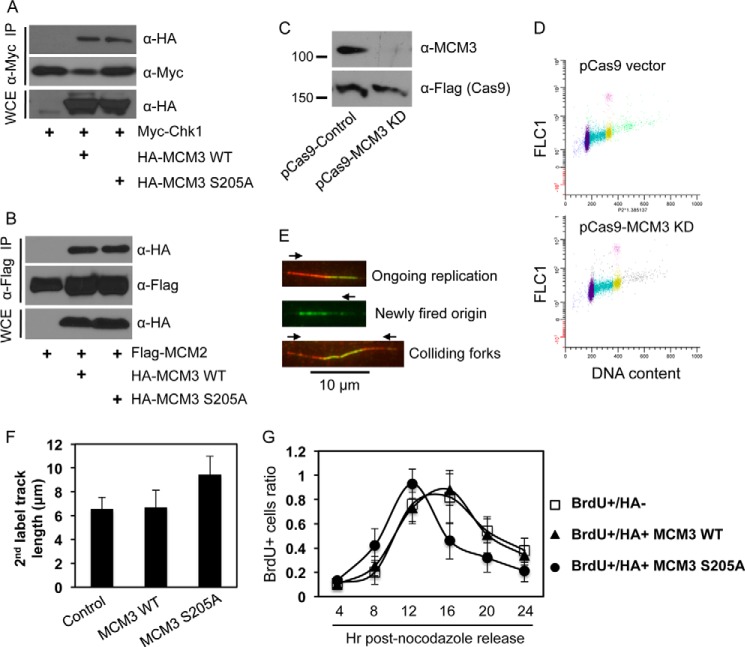
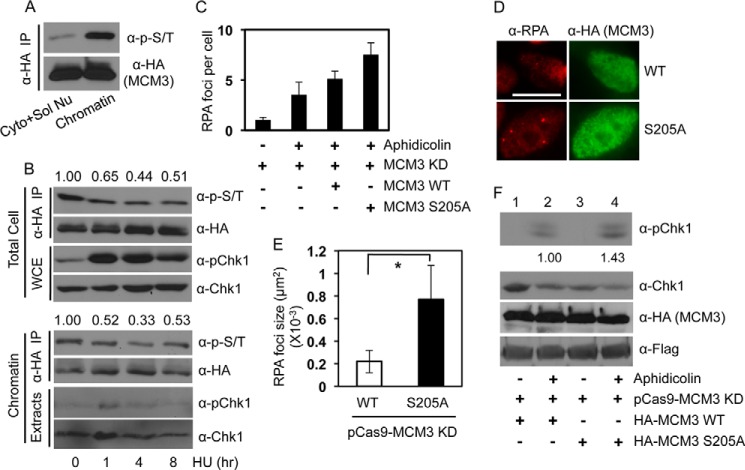
Mechanisms controlling DNA replication and replication checkpoint are critical for the maintenance of genome stability and the prevention or treatment of human cancers. Checkpoint kinase 1 (Chk1) is a key effector protein kinase that regulates the DNA damage response and replication checkpoint. The heterohexameric minichromosome maintenance (MCM) complex is the core component of mammalian DNA helicase and has been implicated in replication checkpoint activation. Here we report that Chk1 phosphorylates the MCM3 subunit of the MCM complex at Ser-205 under normal growth conditions. Mutating the Ser-205 of MCM3 to Ala increased the length of DNA replication track and shortened the S phase duration, indicating that Ser-205 phosphorylation negatively controls normal DNA replication. Upon replicative stress treatment, the inhibitory phosphorylation of MCM3 at Ser-205 was reduced, and this reduction was accompanied with the generation of single strand DNA, the key platform for ataxia telangiectasia mutated and Rad3-related (ATR) activation. As a result, the replication checkpoint is activated. Together, these data provide significant insights into the regulation of both normal DNA replication and replication checkpoint activation through the novel phosphorylation of MCM3 by Chk1.

Keywords: Cell Cycle; DNA Damage; DNA Replication; Phosphorylation; Signal Transduction.


Figures

FIGURE 1.
FIGURE 1.
Phosphorylation of MCM3 by Chk1. A, HEK293T cells were transfected with tagged MCM proteins for 48 h, lysed, IPed with antibodies against the tag, and collected on Protein A/G-agarose beads. The beads were treated with 30 units of CIAP at 37 °C for 1 h, and the in vitro kinase assay was performed in the presence or absence of 10 mm ATP with His-Chk1 as the protein kinase. A GST-tagged Cdc25 fragment (200–250) was used as the positive control. The membrane was probed with an antibody mixture at an equal ratio that can recognize potential Chk1 phosphorylation sites. The same membrane was stripped, cut into small sections based on the protein marker, and reblotted with antibodies against Chk1, the tags for MCMs, or GST. Protein markers are shown on the left. Arrows indicate MCM proteins that were phosphorylated. The asterisk indicates a non-MCM protein that was also phosphorylated. B, HEK293T cells were transfected with HA-MCM3, IPed with anti-HA antibodies, treated or not with CIAP, and blotted with the anti-Ser(P)/Thr(P) mixture. The same membrane was stripped and reblotted with anti-HA antibodies. C, an in vitro kinase assay was performed as in A except that the His-Chk1 protein was pretreated with 500 nm Chk1specific inhibitor (Chk1i) for 2 h on ice. Short and long exposures of the ant-Ser(P)/Thr(P) antibody mixture are illustrated to show the difference in the phospho-MCM3 level between cell cultures and in vitro kinase reaction. The arrow denotes HA-MCM3 protein, and the arrowhead in the anti-Chk1 blot indicates autophosphorylated Chk1 proteins. The asterisk indicates the same non-MCM protein as in A.
FIGURE 2.
FIGURE 2.
MCM3 phosphorylation dependent on Chk1. A, the same in vitro kinase was performed as in Fig. 1A using either Chk1 WT or a kinase-dead (KD) mutant as the kinase. B, HEK293T cells were transfected with HA-MCM3. After 48 h, cells were treated with 1 or 2 μm Chk1 inhibitor for 6 h, lysed, IPed with anti-HA antibodies, and immunoblotted with the anti-Ser(P)/Thr(P) mixture. The same membrane was stripped and reblotted with anti-HA antibodies. Protein expression in whole cell extracts (WCE) was assessed. C, HEK293T cells were treated or not with 1 μm Chk1 inhibitor (Chk1i) for 12 h, lysed, IPed with anti-MCM3 antibodies, and immunoblotted with the indicated antibodies. D, cells treated with 1 μm Chk1 inhibitor for 6 and 12 h were analyzed for the cell cycle profile.
FIGURE 3.
FIGURE 3.
Identification of MCM3 phosphorylation sites. A, schematic diagram of MCM3 FL and four fragments (1–4). AAA+, ATPase domain; NTD, nucleic acid-binding domain. B, HEK293T cells were transfected with HA-MCM3 FL (WT, S205A, or S419A) or fragment 1 (WT or S205A) for 48 h, IPed with anti-HA antibodies, and blotted with the anti-Ser(P)/Thr(P) mixture (left). The same membrane was stripped and reblotted with anti-HA antibodies (right). C, HA-MCM3 WT or S205A mutant was collected from transfected HEK293T cells, treated with CIAP, and used as the substrate for in vitro kinase assay in the presence of [γ-32P]ATP. The SDS-polyacrylamide gel was stained with Coomassie Blue (lower), dried, and exposed to x-ray radiography (upper).
FIGURE 4.
FIGURE 4.
Ser-205 is the major site of MCM3 phosphorylated by Chk1. A, HA-MCM3 WT proteins were collected by IP from HEK293T cells, treated with CIAP, used in the in vitro kinase reaction, and run on SDS-PAGE, and the corresponding band was excised and used for mass spectrometry analyses. Phosphorylation was detected for the peptide 203–222 (one of the following four residues: Tyr-204, Ser-205, Thr-208, and Thr-209). B, HEK293T cells were transfected with HA-MCM3 FL (WT, S205A, or ASDLAA) for 48 h, IPed with anti-HA antibodies, and blotted with the anti-Ser(P)/Thr(P) mixture. The same membrane was stripped and reblotted with anti-HA antibodies.
FIGURE 5.
FIGURE 5.
Ser-205 phosphorylation of MCM3 negatively regulates DNA replication and cell cycle progression. A, HEK293T cells were transfected with Myc-Chk1 and HA-MCM3 WT or S205A, IPed with anti-Myc antibodies, and blotted with anti-HA antibodies. The same membrane was stripped and reblotted with anti-Myc antibodies. B, HEK293T cells were transfected with FLAG-MCM2 and HA-MCM3 WT or S205A, IPed with anti-FLAG antibodies, and blotted with anti-HA antibodies followed by stripping and reprobing with anti-FLAG antibodies. C, U2-OS cells were transfected twice in consecutive days with the CRISPR/Cas9 system that targets the 3′- and 5′-UTRs of human MCM3. 48 h after the second transfection (total 72 h of knockdown), cells were collected and immunoblotted with the indicated antibodies. The Cas9 is expressed as a FLAG fusion protein. D, cell cycle profile of U2-OS cells after 72 h of transfection with the control pCas9 or pCas9-MCM3 knockdown (KD) vector. E, DNA fiber assay. U2-OS cells were transfected twice in consecutive days with the CRISPR/Cas9 vectors targeting both the 3′- and 5′-UTRs of MCM3. During the second transfection, HA-MCM3 WT or S205A vectors were also transfected. 48 h after the second transfection, DNA replication in cells was analyzed by the DNA fiber assay as detailed under “Experimental Procedures.” Red and green represent initial and subsequent replication tracks, respectively, taken under a fluorescence microscope (100× oil lens). F, a DNA fiber assay was carried out in U2-OS cells expressing HA-MCM3 WT or S205A mutant under the background of depletion of endogenous MCM3 by the CRISPR/Cas system. The length of actively replicating DNA tracks was analyzed using NIH ImageJ software and adjusted to a standard scale bar of 10 μm taken under the same condition. A minimum of 100 tracks was measured from duplicated experiments, and the experiment was repeated twice. Data represent median and S.D. (error bars). G, U2-OS cells grown on glass coverslips were co-transfected with the CRISPR/Cas MCM3 knockdown plasmid and HA-MCM3 WT or S205A for 72 h, blocked at the G2/M phase by treatment with 200 ng/ml nocodazole for 20 h, and released into the cell cycle. Cells were pulsed with 40 μm BrdU for 15 min at 4, 8, 12, 16, 20, and 24 h after nocodazole release; fixed; immunostained with antibodies against HA and BrdU; and visualized under a fluorescence microscope. The percentage of BrdU-positive cells in HA-negative or HA-positive populations was determined. Data represent median and S.D. (error bars) from duplicate experiments repeated twice. More than 50 cells were counted in each duplicate. WCE, whole cell extracts.
FIGURE 6.
FIGURE 6.
Roles of MCM3 phosphorylation in replication checkpoint activation. A, HEK293T cells were transfected with HA-MCM3 WT, fractionated into chromatin and non-chromatin fractions, IPed with anti-HA antibodies, and blotted with anti-Ser(P)/Thr(P) antibodies followed by stripping and reprobing with anti-HA antibodies. B, HEK293T cells were transfected with HA-MCM3 for 48 h and treated with 2 mm hydroxyurea (HU) for 0, 1, 4 and 8 h. Whole cell extracts (WCE) or chromatin-enriched fraction were IPed with anti-HA antibodies and blotted with the anti-Ser(P)/Thr(P) mixture. The same membrane was stripped and reblotted with anti-HA antibodies. Phosphorylated Chk1 (pChk1) and total Chk1 in the extracts were also monitored. The band intensity of both the Ser(P)/Thr(P) and the HA blots was analyzed using NIH ImageJ software, and the relative intensity of the Ser(P)/Thr(P) blot after adjusting against the anti-HA blot intensity is shown from three replicates. C, U2-OS cells grown on glass coverslips were co-transfected with the CRISPR/Cas MCM3 knockdown (KD) plasmid and HA-MCM3 WT or S205A for 72 h, treated with 1.5 μg/ml aphidicolin for 1 h, fixed, and immunostained with anti-HA and anti-RPA antibodies. Data represent the number of RPA foci per cell from 30–50 cells in duplicate. D, representative images of RPA foci from C. Scale bar, 10 μm. E, quantitation results of RPA focus size by NIH ImageJ software from cells in D. Data represent median and S.D. (error bars) from 30–50 cells in duplicate. *, p < 0.05. F, U2-OS cells were co-transfected with the CRISPR/Cas MCM3 knockdown plasmid and HA-MCM3 WT or S205A for 72 h and treated with 1.5 μg/ml aphidicolin for 1 h, and protein expression was analyzed. The phospho-Chk1 blot was stripped and reprobed with anti-Chk1 antibodies. The protein band intensity of Chk1 and phospho-Chk1 in lanes 2 and 4 was quantitated using NIH ImageJ software. The relative level of phospho-Chk1 was normalized to that of total Chk1, which shows a ∼40% increase averaged from three blots. Cyto + Sol Nu, cytosol and soluble nuclear proteins.

Similar articles

Cited by

References

    1. Harper J. W., Elledge S. J. (2007) The DNA damage response: ten years after. Mol. Cell 28, 739–745 - PubMed
    1. Jackson S. P., Bartek J. (2009) The DNA-damage response in human biology and disease. Nature 461, 1071–1078 - PMC - PubMed
    1. Zhang Y., Hunter T. (2014) Roles of Chk1 in cell biology and cancer therapy. Int. J. Cancer 134, 1013–1023 - PMC - PubMed
    1. Cimprich K. A., Cortez D. (2008) ATR: an essential regulator of genome integrity. Nat. Rev. Mol. Cell Biol. 9, 616–627 - PMC - PubMed
    1. Liu Q., Guntuku S., Cui X. S., Matsuoka S., Cortez D., Tamai K., Luo G., Carattini-Rivera S., DeMayo F., Bradley A., Donehower L. A., Elledge S. J. (2000) Chk1 is an essential kinase that is regulated by Atr and required for the G2/M DNA damage checkpoint. Genes Dev. 14, 1448–1459 - PMC - PubMed

Publication types

MeSH terms