. 2015 May 1;290(18):11393-402.
doi: 10.1074/jbc.M115.637132. Epub 2015 Mar 25.

Pathogenic mutations differentially affect the catalytic activities of the human B12-processing chaperone CblC and increase futile redox cycling

Affiliations

Pathogenic mutations differentially affect the catalytic activities of the human B12-processing chaperone CblC and increase futile redox cycling

Carmen Gherasim et al. J Biol Chem. .

Abstract

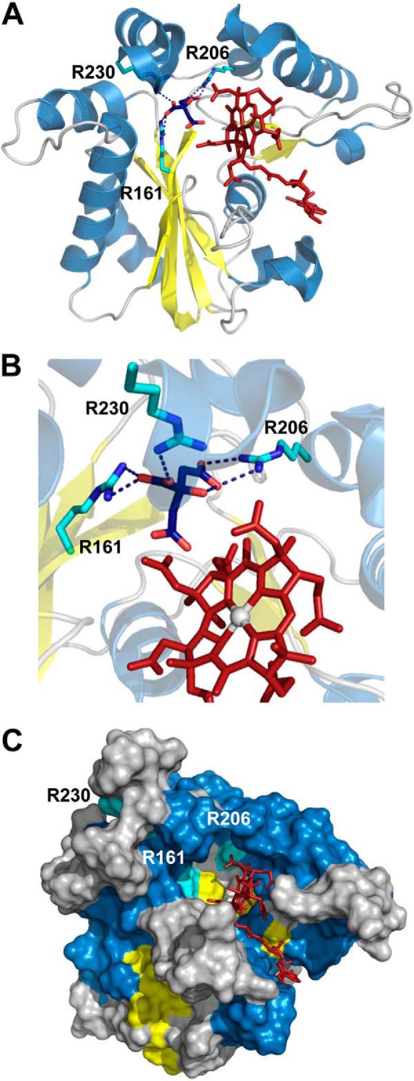
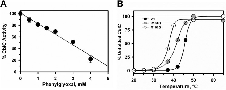
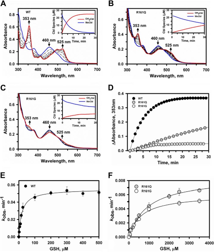
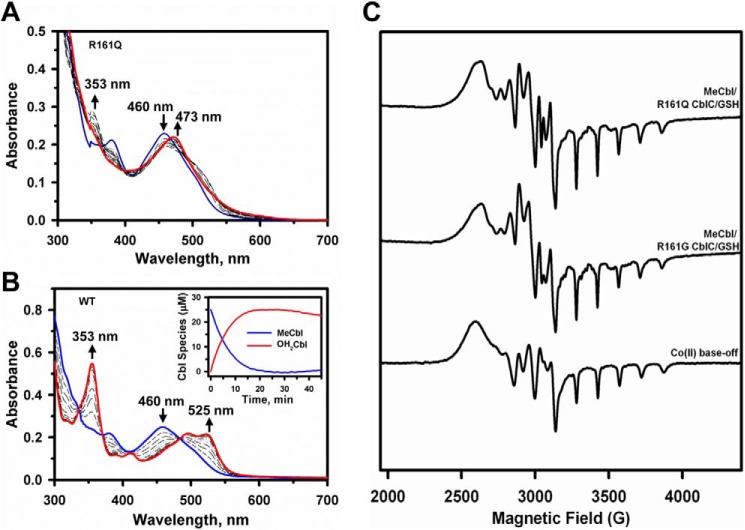
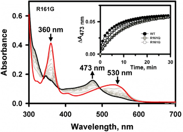
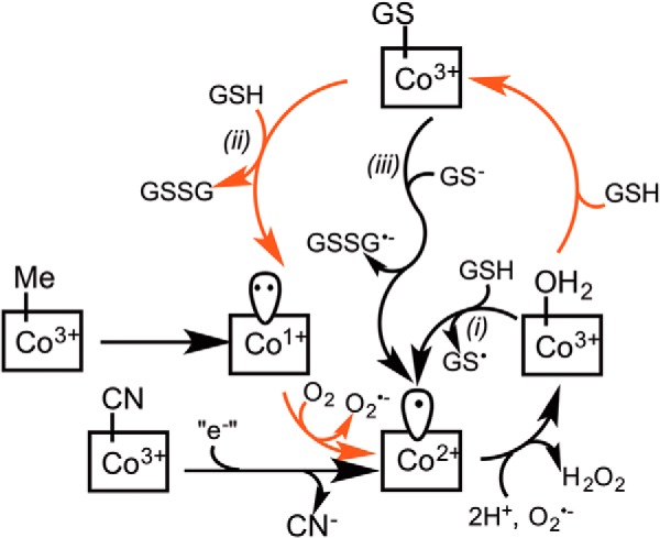
Human CblC catalyzes the elimination of the upper axial ligand in cobalamin or B12 derivatives entering the cell from circulation. This processing step is critical for assimilation of dietary cobalamin into the active cofactor forms that support the B12-dependent enzymes, methionine synthase and methylmalonyl-CoA mutase. Using a modified nitroreductase scaffold tailored to bind cobalamin and glutathione, CblC exhibits versatility in the mechanism by which it removes cyano versus alkyl ligands in cobalamin. In this study, we have characterized the effects of two pathogenic missense mutations at the same residue, R161G and R161Q, which are associated with early and late onset of the CblC disorder, respectively. We find that the R161Q and R161G CblC mutants display lower protein stability and decreased dealkylation but not decyanation activity, suggesting that cyanocobalamin might be therapeutically useful for patients carrying mutations at Arg-161. The mutant proteins also exhibit impaired glutathione binding. In the presence of physiologically relevant glutathione concentrations, stabilization of the cob(II)alamin derivative is observed, which occurs at the expense of increased oxidation of glutathione. Futile redox cycling, which is suppressed in wild-type human CblC, explains the reported increase in oxidative stress levels associated with the CblC disorder.

Keywords: Adenosylcobalamin (AdoCbl); Chaperone; Cofactor; Enzyme Kinetics; Oxidation-Reduction (Redox); Trafficking.


Figures

FIGURE 1.
FIGURE 1.
Structure of human CblC with AdoCbl bound (Protein Data Bank (PDB) 3SOM) showing the locations of Arg-161, Arg-206 and Arg-230 predicted to be involved in GSH-binding. A, cartoon representation of CblC structure (PDB: 3SOM) with helixes, β-sheets, and loops colored in blue, yellow, and gray, respectively. AdoCbl is shown in stick representation in red, and Arg-161, Arg-206, and Arg-230 are shown in cyan. Citrate bound to the active site is represented in blue. B, close-up of the hydrogen-bonding interactions between Arg-161, Arg-206, and Arg-230 and citrate bound at the active site. C, surface representation of the CblC structure indicating accessibility of the upper and lower faces of the corrin ring in the absence of bound GSH.
FIGURE 2.
FIGURE 2.
Effect of arginines on the activity and stability of CblC. A, phenylglyoxal inhibits dealkylation of MeCbl by CblC. CblC (30 μm) and MeCbl (20 μm) in Buffer A were incubated with 10 mm GSH and increasing concentrations of phenylglyoxal (0–4 mm) for 30 min, and its effect on the demethylation activity was determined as described under “Experimental Procedures.” Plots were normalized to denote maximum activity in the absence of phenylglyoxal. The data represent the mean ± S.D. of three independent experiments. B, the effect of R161Q/G substitutions on the stability of CblC as monitored at 600 nm. Plots for the temperature-dependent unfolding of R161Q (gray circles) and R161G (open circles) mutants are compared with wild-type (black circles) CblC. The plots were normalized to the starting absorbance at 600 nm representing folded protein. The Tm values were determined as described under “Experimental Procedures.”
FIGURE 3.
FIGURE 3.
Dealkylation of MeCbl by wild-type and R161Q/G CblC. A–C, time-dependent changes in the UV-visible absorption spectra of MeCbl (25 μm) bound to wild-type (A), R161Q (B), or R161G (C) CblC (30 μm) in Buffer A following the addition of 1 mm GSH under aerobic conditions, at 25 °C. The initial CblC-bound MeCbl spectrum has an absorption maxima at 460 nm (blue trace), whereas the product spectra recorded at 30 min shows features characteristic for OH2Cbl at 353 and 525 nm (red traces). The insets in in A–C show the concentration of the MeCbl consumed and OH2Cbl formed during the reactions. D, comparison of the MeCbl dealkylation rates for wild-type (black circles), R161Q (gray circles), and R161G CblC (open circles). E and F, dependence of the initial rate of dealkylation of MeCbl bound to wild-type (E) or R161Q/G (F) CblC on GSH concentration. Aerobic reaction mixtures containing 30 μm CblC, 25 μm MeCbl, and 0–5 mm GSH in Buffer A were monitored at 25 °C at 353 nm. The data are the mean ± S.D. of at least three independent experiments.
FIGURE 4.
FIGURE 4.
Stabilization of cob(II)alamin by R161Q/G CblC at high GSH concentrations. UV-visible spectra were recorded to monitor dealkylation of MeCbl. A and B, the reaction mixtures contained R161Q CblC (A) or wild-type CblC (30 μm) and MeCbl (25 μm) (B) in aerobic Buffer A at 25 °C. The initial spectra are shown in blue, and spectra were recorded every 3 min following the addition of 10 mm GSH (only a few spectra are shown for clarity). The spectral changes were consistent with formation of cob(II)alamin with an absorption maximum at 473 nm with R161Q CblC (red trace, A) and OH2Cbl with absorption maxima at 353 and 525 nm with wild-type CblC (red trace, B). C, EPR spectra of authentic cob(II)alamin (lower) generated as described under “Experimental Procedures” compared with cob(II)alamin formed during dealkylation of MeCbl catalyzed by R161Q (upper) and R161G (middle) CblC. The samples were prepared as described under “Experimental Procedures.”
FIGURE 5.
FIGURE 5.
Decyanation kinetics of wild-type and mutant CblCs. Shown are changes in the UV-visible absorption spectrum of CNCbl (20 μm) bound to R161G CblC (50 μm) in Buffer A at 25 °C following addition to a mixture of methionine synthase reductase/NADPH (4 μm/200 μm) under anaerobic conditions. The initial spectrum is in red, selected traces over the reaction course are in gray, and the final spectrum at 60 min is in black. The inset shows the reaction kinetics monitored at 473 nm for wild-type (black circles), R161Q (gray circles), and R161G (open circles) CblC.
FIGURE 6.
FIGURE 6.
GSH-dependent reduction of OH2Cbl by wild-type and R161Q/G CblC. A and B, changes in the UV-visible absorption spectra of OH2Cbl (20 μm) bound to R161G CblC (50 μm) in Buffer A were monitored at 25 °C under aerobic (A) and anaerobic conditions (B). Reactions were initiated by the addition of 10 mm GSH. The initial spectra are in red, selected traces over the reaction course time are in gray, and the final spectra at 60 min are in black. Following the addition of GSH to the aerobic sample (A), the absorption maximum shifts from 525 to 516 nm (red dashed trace) before cob(II)alamin formation (473 nm maximum) is observed. The insets in A and B show the reaction kinetics monitored at 475 nm for wild-type (black circles), R161Q (gray circles), and R161G (open circles) CblC. The slight decrease in absorbance at 475 nm over time observed with the more active R161G CblC suggests slow oxidation of cob(II)alamin to OH2Cbl. C, HPLC analysis of GSSG formed after 60 min in the assay mixtures during the reaction shown in A. The data represent the mean ± S.D. of three independent experiments. D, oxygen consumption kinetics associated with GSH-dependent reduction of OH2Cbl in the presence of wild-type (black circles), R161Q (gray circles), and R161G (open circles) CblC.
FIGURE 7.
FIGURE 7.
Alternative mechanisms for redox cycling and cob(II)alamin stabilization by the R161Q/G mutants. Demethylation of MeCbl by CblC leads to cob(I)alamin formation, which is labile and is readily oxidized to form cob(II)alamin, and subsequently, to OH2Cbl, which is stabilized by wild-type CblC. Enhanced formation of GSSG accompanying cob(II)alamin stabilization by the R161 mutants could occur by one of three routes denoted i–iii. In path i, OH2Cbl is reduced by GSH to cob(II)alamin. In paths ii and iii, OH2Cbl is initially converted to GSCbl. The latter is either displaced by a second mole of GSH (path ii) or reduced, leading to cob(II)alamin (path iii). The GS radical formed in path i is rapidly quenched by GSH, giving GSSG•−, which is also formed in path iii, and the GSSG•− radical reacts rapidly with O2 to form O2, further enhancing ROS production. The reactions that are predicted to be accelerated in the Arg-161 mutants are depicted by red arrows.

References

    1. Banerjee R., Ragsdale S. W. (2003) The many faces of vitamin B12: catalysis by cobalamin-dependent enzymes. Annu. Rev. Biochem. 72, 209–247 - PubMed
    1. Banerjee R. (2001) Radical peregrinations catalyzed by coenzyme B12-dependent enzymes. Biochemistry 40, 6191–6198 - PubMed
    1. Banerjee R., Gherasim C., Padovani D. (2009) The tinker, tailor, soldier in intracellular B12 trafficking. Curr. Opin. Chem. Biol. 13, 484–491 - PMC - PubMed
    1. Gherasim C., Lofgren M., Banerjee R. (2013) Navigating the B12 road: assimilation, delivery and disorders of cobalamin. J. Biol. Chem. 288, 13186–13193 - PMC - PubMed
    1. Watkins D., Rosenblatt D. S. (2012) Update and new concepts in vitamin responsive disorders of folate transport and metabolism. J. Inherit. Metab. Dis. 35, 665–670 - PubMed

Publication types

MeSH terms

Associated data

LinkOut - more resources