. 2015 May 8;290(19):12237-46.
doi: 10.1074/jbc.M115.643098. Epub 2015 Mar 25.

Self-cleavage of the Pseudomonas aeruginosa Cell-surface Signaling Anti-sigma Factor FoxR Occurs through an N-O Acyl Rearrangement

Affiliations

Self-cleavage of the Pseudomonas aeruginosa Cell-surface Signaling Anti-sigma Factor FoxR Occurs through an N-O Acyl Rearrangement

Karlijn C Bastiaansen et al. J Biol Chem. .

Abstract

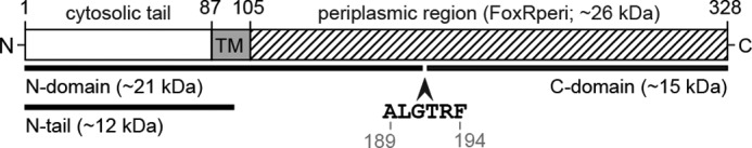
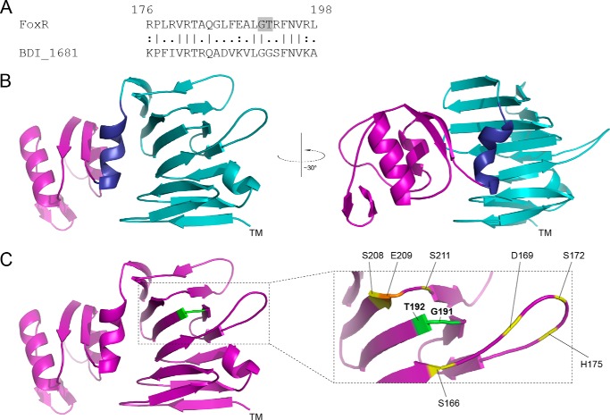
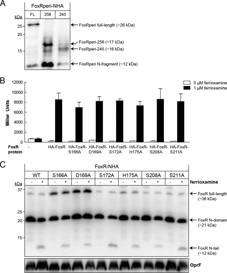
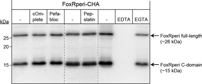
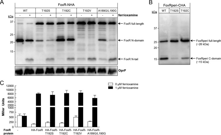
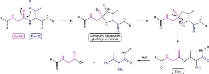
The Fox system of Pseudomonas aeruginosa is a cell-surface signaling (CSS) pathway employed by the bacterium to sense and respond to the presence of the heterologous siderophore ferrioxamine in the environment. This regulatory pathway controls the transcription of the foxA ferrioxamine receptor gene through the extracytoplasmic function sigma factor σ(FoxI). In the absence of ferrioxamine, the activity of σ(FoxI) is inhibited by the transmembrane anti-sigma factor FoxR. Upon binding of ferrioxamine by the FoxA receptor, FoxR is processed by a complex proteolytic cascade leading to the release and activation of σ(FoxI). Interestingly, we have recently shown that FoxR undergoes self-cleavage between the periplasmic Gly-191 and Thr-192 residues independent of the perception of ferrioxamine. This autoproteolytic event, which is widespread among CSS anti-sigma factors, produces two distinct domains that interact and function together to transduce the presence of the signal. In this work, we provide evidence that the self-cleavage of FoxR is not an enzyme-dependent process but is induced by an N-O acyl rearrangement. Mutation analysis showed that the nucleophilic side chain of the Thr-192 residue at +1 of the cleavage site is required for an attack on the preceding Gly-191, after which the resulting ester bond is likely hydrolyzed. Because the cleavage site is well preserved and the hydrolysis of periplasmic CSS anti-sigma factors is widely observed, we hypothesize that cleavage via an N-O acyl rearrangement is a conserved feature of these proteins.

Keywords: Cell Signaling; Gene Regulation; Iron; Post-translational Modification (PTM); Proteolysis; Pseudomonas aeruginosa (P. aeruginosa); Siderophore; Signal Transduction.


Figures

FIGURE 1.
FIGURE 1.
Schematic representation of the P. aeruginosa FoxR protein. The P. aeruginosa FoxR protein has been drawn to scale, and the cytosolic, transmembrane, and periplasmic (FoxRperi) regions of the protein are detailed. The site where the self-cleavage of FoxR occurs (between Gly-191 and Thr-192; GT) and surrounding residues have been indicated. The numbers indicate amino acid positions in the FoxR protein. The N- and C-domains resulting from self-cleavage are illustrated. The exact cleavage sites of the Prc and RseP proteases are unknown.
FIGURE 2.
FIGURE 2.
Structural modeling of PaFoxR. A, a pair-wise alignment of the P. aeruginosa FoxR protein (residues 176–198) is shown with its homologue BDI_1681 of P. distasonis. The GT cleavage site in FoxR is shaded. B, a cartoon representation of the structural model of the periplasmic region of FoxR (FoxRperi) created using the Phyre program (32) is shown. The location of the transmembrane segment is indicated by TM. The C terminus of FoxRperi (residues 256–328) has been colored purple, and the N terminus of FoxRperi (residues 114–245) is in light blue. The helix separating these distinct regions (residues 246–255) is shown in dark blue. C, the residues flanking the self-cleavage site (Gly-191 and Thr-192) have been colored green. Residues that could be part of a putative proteolytic active site (Ser, etc.) are indicated in yellow, and Glu-209 is shown in orange (see text).
FIGURE 3.
FIGURE 3.
Mutational analysis of putative active site residues of PaFoxR. A, the FoxRperi protein with an N-terminal HA tag (FoxRperi-NHA-FL; amino acids 107–328) was synthesized using the PURExpress® in vitro transcription/translation system. In addition, a FoxRperi-NHA truncate lacking the complete C terminus (FoxRperi-NHA-256; amino acids 107–256) and a truncate also lacking the α-helix (FoxRperi-NHA-245; amino acids 107–245) were produced. Reactions were analyzed by anti-HA immunoblot. B, β-galactosidase activity of the P. aeruginosa PAO1 pvdF ΔfoxR mutant bearing the pMPR8b plasmid (foxA::lacZ transcriptional fusion) and the pMMB67EH (empty), pMMB/HA-FoxR (WT), pMMB/HA-FoxR-S166A, pMMB/HA-FoxR-D169A, pMMB/HA-FoxR-S172A, pMMB/HA-FoxR-H175A, pMMB/HA-FoxR-S208A, or pMMB/HA-FoxR-S211A plasmid expressing the corresponding N-terminally HA-tagged FoxR protein. Bacteria were grown in iron-restricted CAS medium without or with 1 μm ferrioxamine. C, Western blot analysis of the P. aeruginosa pvdF ΔfoxR mutant bearing the pMMB/HA-FoxR (WT) plasmid or one of its derivative plasmids expressing one of the FoxR-S166A, -D169A, -S172A, H175A, or -S211A mutant variants. Bacteria were grown in iron-restricted medium with 1 mm IPTG in the absence (−) or presence (+) of 1 μm ferrioxamine. Proteins were detected using a monoclonal anti-HA tag antibody. As a loading control, a monoclonal antibody against the OprF protein was used. The positions of the molecular size marker (in kDa) and the FoxR protein fragments are indicated.
FIGURE 4.
FIGURE 4.
Effect of protease inhibitors on PaFoxR self-cleavage. FoxRperi with a C-terminal HA tag (FoxRperi-CHA) was produced using the PURExpress® in vitro transcription/translation system in the presence of protease inhibitors. Subsequently, the reaction products were analyzed by Western blot using a monoclonal anti-HA antibody. The positions of the molecular size marker (in kDa) and the FoxR protein fragments are indicated.
FIGURE 5.
FIGURE 5.
Potential role of an N-O acyl rearrangement in PaFoxR self-cleavage. A, anti-HA tag immunoblot of P. aeruginosa pvdF ΔfoxR carrying the pMMB/HA-FoxR (WT), pMMB/HA-FoxR-T192S, pMMB/HA-FoxR-T192C, pMMB/HA-FoxR-T192V, or pMMB/HA-FoxR-A189G/L190G plasmid. Bacteria were grown under iron-restricted conditions in the presence of 1 mm IPTG without (−) and with (+) 1 μm ferrioxamine. B, Western blot of FoxRperi-CHA incorporating either the T192S or T192V mutation during expression in the PURExpress® system. Proteins were detected using a monoclonal anti-HA tag antibody. The positions of the molecular size marker (in kDa) and the FoxR protein fragments in A and B are indicated. As a loading control in A, a monoclonal antibody against the OprF protein was used. C, β-galactosidase activity of the P. aeruginosa PAO1 pvdF ΔfoxR mutant bearing the pMPR8b plasmid (foxA::lacZ transcriptional fusion) and the pMMB67EH (empty), pMMB/HA-FoxR (WT), pMMB/HA-FoxR-T192S, pMMB/HA-FoxR-T192C, pMMB/HA-FoxR-T192V, or pMMB/HA-FoxR-A189G/L190G plasmid. Bacteria were grown in iron-restricted medium in the absence or presence of 1 μm ferrioxamine.
FIGURE 6.
FIGURE 6.
Proposed sequence for self-cleavage of PaFoxR. An initial N-O acyl rearrangement is followed by ester hydrolysis. The two R labels indicate the remainders of the protein chain. The cores of Gly-191 and Thr-192 have been colored for clarity.

Similar articles

Cited by

References

    1. Llamas M. A., Sparrius M., Kloet R., Jiménez C. R., Vandenbroucke-Grauls C., Bitter W. (2006) The heterologous siderophores ferrioxamine B and ferrichrome activate signaling pathways in Pseudomonas aeruginosa. J. Bacteriol. 188, 1882–1891 - PMC - PubMed
    1. Ratledge C., Dover L. G. (2000) Iron metabolism in pathogenic bacteria. Annu. Rev. Microbiol. 54, 881–941 - PubMed
    1. Wandersman C., Delepelaire P. (2004) Bacterial iron sources: from siderophores to hemophores. Annu. Rev. Microbiol. 58, 611–647 - PubMed
    1. Noinaj N., Guillier M., Barnard T. J., Buchanan S. K. (2010) TonB-dependent transporters: regulation, structure, and function. Annu. Rev. Microbiol. 64, 43–60 - PMC - PubMed
    1. Poole K., McKay G. A. (2003) Iron acquisition and its control in Pseudomonas aeruginosa: many roads lead to Rome. Front. Biosci. 8, d661–d686 - PubMed

Publication types

MeSH terms

LinkOut - more resources