Review
. 2015 Aug 15;593(16):3447-62.
doi: 10.1113/JP270254.

The regulation of neuronal mitochondrial metabolism by calcium

Affiliations
Review

The regulation of neuronal mitochondrial metabolism by calcium

I Llorente-Folch et al. J Physiol. .

Abstract

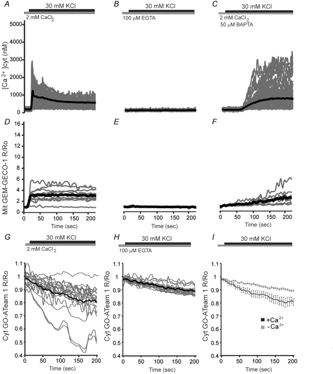
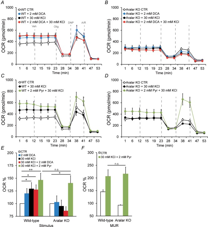
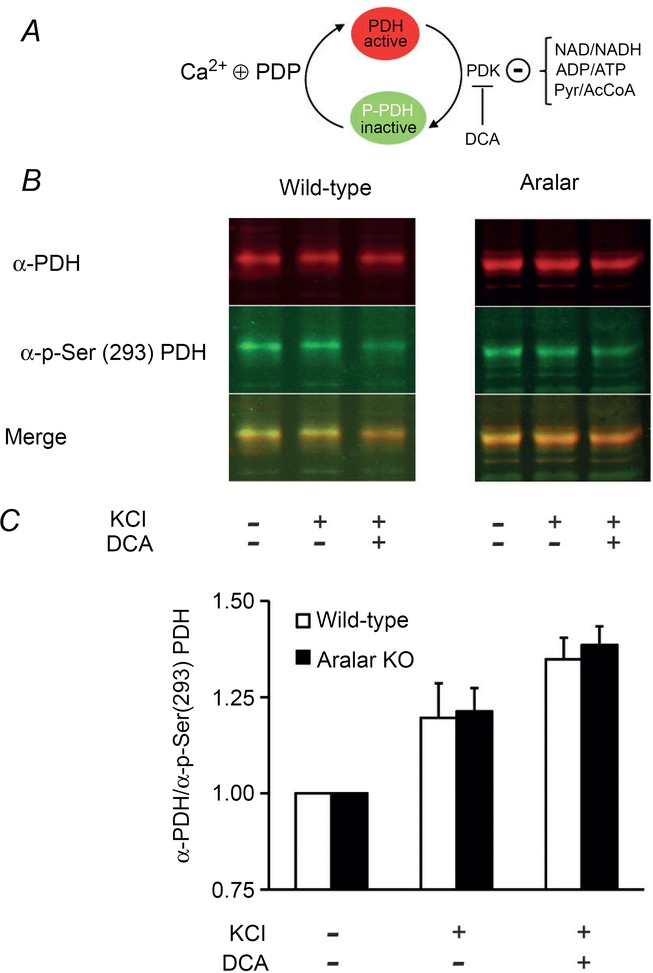
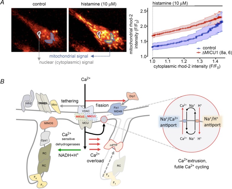
Calcium signalling is fundamental to the function of the nervous system, in association with changes in ionic gradients across the membrane. Although restoring ionic gradients is energetically costly, a rise in intracellular Ca(2+) acts through multiple pathways to increase ATP synthesis, matching energy supply to demand. Increasing cytosolic Ca(2+) stimulates metabolite transfer across the inner mitochondrial membrane through activation of Ca(2+) -regulated mitochondrial carriers, whereas an increase in matrix Ca(2+) stimulates the citric acid cycle and ATP synthase. The aspartate-glutamate exchanger Aralar/AGC1 (Slc25a12), a component of the malate-aspartate shuttle (MAS), is stimulated by modest increases in cytosolic Ca(2+) and upregulates respiration in cortical neurons by enhancing pyruvate supply into mitochondria. Failure to increase respiration in response to small (carbachol) and moderate (K(+) -depolarization) workloads and blunted stimulation of respiration in response to high workloads (veratridine) in Aralar/AGC1 knockout neurons reflect impaired MAS activity and limited mitochondrial pyruvate supply. In response to large workloads (veratridine), acute stimulation of respiration occurs in the absence of MAS through Ca(2+) influx through the mitochondrial calcium uniporter (MCU) and a rise in matrix [Ca(2+) ]. Although the physiological importance of the MCU complex in work-induced stimulation of respiration of CNS neurons is not yet clarified, abnormal mitochondrial Ca(2+) signalling causes pathology. Indeed, loss of function mutations in MICU1, a regulator of MCU complex, are associated with neuromuscular disease. In patient-derived MICU1 deficient fibroblasts, resting matrix Ca(2+) is increased and mitochondria fragmented. Thus, the fine tuning of Ca(2+) signals plays a key role in shaping mitochondrial bioenergetics.


Figures

Figure 1
Figure 1. Schematic representation of Ca2+regulation of mitochondrial respiration
Tricarboxylic acid cycle enzymes are highly sensitive to changes in [Ca2+], which presumably binds directly to isocitrate dehydrogenase (IDH), α-ketoglutarate dehydrogenase (α-KGDH), whereas PDH is activated by the Ca2+-sensitive pyruvate dehydrogenase phosphatase. Complex IV and complex III may also be regulated by intramitochondrial Ca2+. Matrix Ca2+ may also regulate OXPHOS through an effect on the ANT and on the F1Fo-ATP synthase. Extramitochondrial Ca2+ activates Aralar/AGC1-MAS activity and SCaMC-3. P-PDH, phosphorilated pyruvate dehydrogenase; PDK, pyruvate dehydrogenase kinase; PDP, pyruvate dehydrogenase phosphatase; Pyr, pyruvate; AcCoA, acetyl coenzyme A; TCA: tricarboxylic acid cycle; NHX, Na+/H+ exchanger;
Figure 2
Figure 2. Changes of cytosolic and mitochondrial
Ca2+ and cytosolic ATP in cortical neurons in response to potassiumChanges in [Ca2+]cyt, in Fura-2 loaded neurons (AC) or [Ca2+]mit, in neurons transfected with Mit GEM-GECO1 probe (DF) obtained by stimulation with 30 mm KCl in 2 mm Ca2+ medium (A and D), Ca2+ free medium in the presence of the Ca2+ chelator EGTA (B and E), or in 50 μm BAPTA-AM preloaded neurons, an intracellular Ca2+ chelator that preserves the workload preventing Ca2+ signalling, in 2 mm Ca2+ medium (C and F). Recordings from at least 60 cells per condition and two independent experiments were used for cytosolic Ca2+ imaging and a minimum of 15 cells and eight independent experiments were used for mitochondrial Ca2+ imaging. Individual cell recordings (in grey) and average (thick black trace) were shown. GI, cytosolic ATP levels after a switch from HCSS medium to isosmotic high K+ medium in which 30 mm NaCl was replaced by 30 mm KCl either in 2 mm Ca2+ medium (G) or 100 μm EGTA medium (H). Comparison between the two previously mentioned conditions is shown in (I). The drop in ATP values with respect to basal levels 200 s after high K+ stimulation was 18.6 ± 0.3% in the presence and 9 ± 0.1% in the absence of Ca2+ (**P = 0.009, two-tailed unpaired Student's t test). Data are expressed as the mean ± SEM (modified from Llorente-Folch et al. 2013).
Figure 3
Figure 3. OCR responses to potassium in Aralar/AGC1-deficient neurons
Cellular OCR was measured using a Seahorse XF24 Extracellular Flux Analyser (Seahorse Bioscience, North Billerica, MA, USA) (Qian and Van Houten, 2010). Cortical neurons were plated in XF24 V7 cell culture at 1.0 × 10⁵ cells/well and incubated for 9–10 days in a 37ºC, 5% CO2 incubator in serum-free B27-supplemented neurobasal medium with high levels of glucose. To study OCR, cells were equilibrated for 1 h in 2.5 mm glucose HCSS in the presence of 2 mm CaCl2. Next, neurons were either maintained in the same medium or stimulated with 30 mm KCl in 2.5 mm glucose in Ca2+-containing isosmotic HCSS medium in which 30 mm NaCl was replaced by 30 mm KCl at the start of the respirometry experiments. Calibration of respiration took place after the vehicle (veh) injection in port A. Substrates were prepared in the same medium in which the experiment was conducted and were injected from the reagent ports automatically to the wells at the times indicated. Mitochondrial function in neurons was determined through sequential addition of 6 μm oligomycin (Olig), 0.5 mm 2,4-dinitrophenol (DNP) and 1 μm antimycin/1 μm rotenone (A/R). This allowed the determination of basal oxygen consumption (BS), oxygen consumption linked to ATP synthesis (ATP), non-ATP linked oxygen consumption (leak), mitochondrial uncoupled respiration (MUR) and non-mitochondrial oxygen consumption (NM) (Qian and Van Houten, ; Brand and Nicholls, 2011). Respiratory profiles are shown for control (A) and Aralar/AGC1-deficient neurons (B) upon K+ stimulation, in neurons pre-treated or not with 2 mm DCA for 1 h, in the presence of 2 mm Ca2+. Respiratory profiles are shown for control (C) and Aralar/AGC1-deficient neurons (D) upon K+ stimulation, with or without the addition of 2 mm pyruvate just before starting the experiment, in the presence of 2 mm Ca2+. Stimulation of mitochondrial respiration (E) and maximal uncoupled respiration (MUR) (F) is shown upon K+ stimulation after 2 mm DCA pre-teatment or 2 mm pyruvate addition. Data correspond to four or five and two to four independent experiments in wild-type and Aralar/AGC1 KO cultured neurons, respectively (one-way ANOVA, *P ≤ 0.05; **P ≤ 0.01). KCl, isosmotic high K+, 30 mm; Pyr, pyruvate, 2 mm.
Figure 4
Figure 4. Pyruvate dehydrogenase complex dynamics after K+- depolarization in wild-type and Aralar/AGC1 KO primary cortical neurons
A, scheme depicting the complex dynamics of pyruvate dehydrogenase. PDH-E1 subunit is active in its dephosphorylated state. Pyruvate dehydrogenase kinase (PDK), whose activity is negatively controlled by NAD/NADH, ADP/ATP and Pyr/AcCoA ratios, phosphorylates the enzyme in the Ser(293) residue, inactivating the complex. On the other hand, pyruvate dehydrogenase phosphatase (PDP), which is positively regulated by intramitochondrial Ca2+, dephosphorylates PDH-E1, recovering the active form. DCA inhibits PDK, favouring the active form of PDH. B, representative western blot against PDH (anti-PDH subunit E1, mouse monoclonal antibody, dilution 1:5000; Invitrogen, Carlsbad, CA, USA) and p-Ser(293) PDH (anti-PDH, rabbit polyclonal antibody, dilution 1:2000; Novus Biologicals, Littleton, CO, USA) and IRDye secondary antibodies optimized for use with Oddysey (800 CW goat anti-rabbit IgG and 680 RD goat anti-mouse IgG, dilution 1:50000; Li-Cor, Lincoln, NE, USA). Neurons were obtained under control conditions, or after 5 min of exposure to isosmotic 30 mm KCl, or under these same conditions prior to pre-treatment for 1 h with 2 mm DCA. Merged image combines both green and red fluorescence to denote the PDH/phosphorylated pyruvate dehydrogenase (P-PDH) ratio. C, western-blotting quantification expressed as the fold increase in anti-PDH/anti-p-Ser(293) PDH ratio compared to control. Data correspond to three to five independent experiments in wild-type and Aralar/AGC1 KO cultured neurons, respectively (Student's t test, *P < 0.05). Pyr, pyruvate; AcCoA, acetyl coenzyme A; KCl: isosmotic high K+, 30 mm.
Figure 5
Figure 5. Consequences of MICU1/2 loss on mitochondrial Ca2+ homeostasis, shape and metabolic function
A, relationship between cytosolic and mitochondrial [Ca2+] during evoked Ca2+ signals. MICU1/2 defective cells have an increased basal mitochondrial Ca2+ load, which follows linearly the increase in cytoplasmic [Ca2+] (reproduced with permission from Logan et al. 2014). B, increased mitochondrial Ca2+ load can activate mitochondrial metabolism by stimulating Ca2+-dependent dehydrogenases of the mitochondrial matrix. On the other hand, chronic Ca2+ load in the mitochondrion might also have also a metabolic cost by resulting in futile Ca2+ cycling (inset) and opening of the mitochondrial permeability transition pore (mPTP), both leading to depolarization. In addition, we observed increased mitochondrial fission, which might impact on the metabolic capacity of the organelle. In the human fibroblast model of the disease, no changes in ER mitochondria tethering were observed. mfn2, mitofusin 2; ERMES, ER-mitochondria encounter structures; Miro, mitochondrial Rho GTPase; MINOS: MINOS/MitOS/MICOS complexes; RC, respiratory supercomplexes; F1/FO, F1FO ATPase.

References

    1. Amigo I, Traba J, Gonzalez-Barroso MM, Rueda CB, Fernandez M, Rial E, Sanchez A, Satrustegui J, del Arco A. Glucagon regulation of oxidative phosphorylation requires an increase in matrix adenine nucleotide content through Ca2+-activation of the mitochondrial ATP-Mg/Pi carrier SCaMC-3. J Biol Chem. 2013;288:7791–7802. - PMC - PubMed
    1. Aprille JR. Regulation of the mitochondrial adenine nucleotide pool size in liver: mechanism and metabolic role. FASEB J. 1988;2:2547–2556. - PubMed
    1. Aprille JR. Mechanism and regulation of the mitochondrial ATP-Mg/P(i) carrier. J Bioenerg Biomembr. 1993;25:473–481. - PubMed
    1. Asimakis GK, Aprille JR. In vitro alteration of the size of the liver mitochondrial adenine nucleotide pool: correlation with respiratory functions. Arch Biochem Biophys. 1980;203:307–316. - PubMed
    1. Attwell D, Laughlin SB. An energy budget for signaling in the grey matter of the brain. J Cereb Blood Flow Metab. 2001;21:1133–1145. - PubMed

Substances

LinkOut - more resources