. 2015 Jul;4(7):1039-50.
doi: 10.1002/cam4.445. Epub 2015 Mar 23.

An estrogen-induced endometrial hyperplasia mouse model recapitulating human disease progression and genetic aberrations

Affiliations

An estrogen-induced endometrial hyperplasia mouse model recapitulating human disease progression and genetic aberrations

Chieh-Hsiang Yang et al. Cancer Med. 2015 Jul.

Abstract

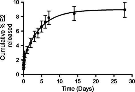
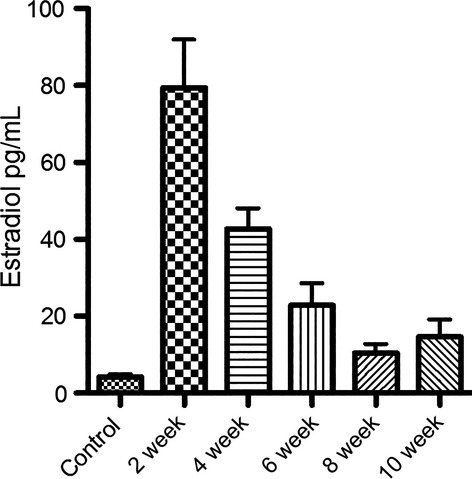
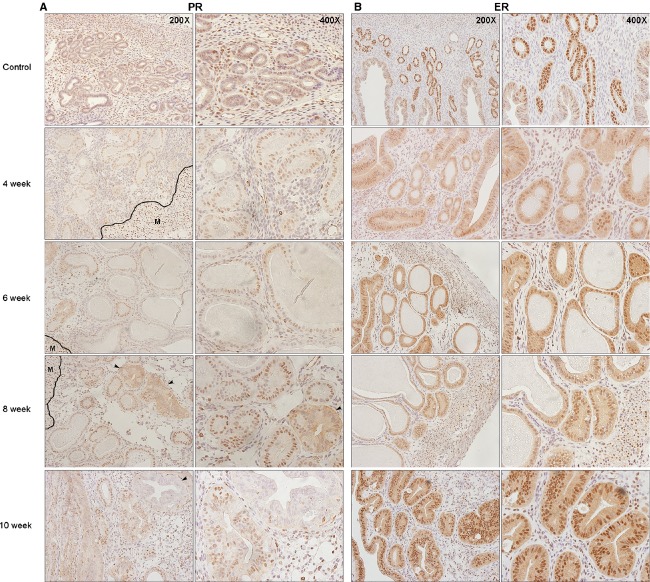
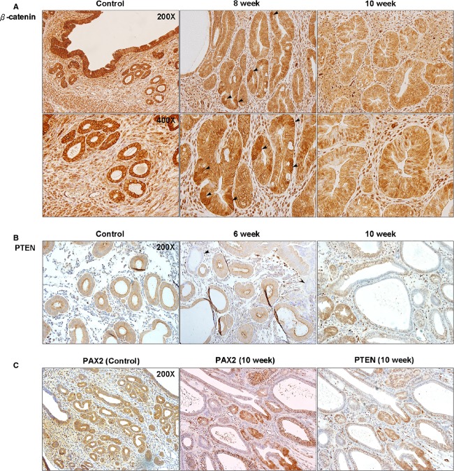
Endometrial hyperplasia (EH) is a condition originating from uterine endometrial glands undergoing disordered proliferation including the risk to progress to endometrial adenocarcinoma. In recent years, a steady increase in EH cases among younger women of reproductive age accentuates the demand of therapeutic alternatives, which emphasizes that an improved disease model for therapeutic agents evaluation is concurrently desired. Here, a new hormone-induced EH mouse model was developed using a subcutaneous estradiol (E2)-sustained releasing pellet, which elevates the serum E2 level in mice, closely mimicking the effect known as estrogen dominance with underlying, pathological E2 levels in patients. The onset and progression of EH generated within this model recapitulate a clinically relevant, pathological transformation, beginning with disordered proliferation developing to simple EH, advancing to atypical EH, and then progressing to precancerous stages, all following a chronologic manner. Although a general increase in nuclear progesterone receptor (PR) expression occurred after E2 expression, a total loss in PR was noted in some endometrial glands as disease advanced to simple EH. Furthermore, estrogen receptor (ER) expression in the nucleus of endometrial cells was reduced in disordered proliferation and increased when EH progressed to atypical EH and precancerous stages. This EH model also resembles other pathological patterns found in human disease such as leukocytic infiltration, genetic aberrations in β-catenin, and joint phosphatase and tensin homolog/paired box gene 2 (PTEN/PAX2) silencing. In summary, this new and comprehensively characterized EH model is cost-effective, easily reproducible, and may serve as a tool for preclinical testing of therapeutic agents and facilitate further investigation of EH.

Keywords: Endometrial cancer; endometrial hyperplasia; estrogen; mouse model.


Figures

Figure 1
Figure 1
In vitro release kinetics of E2 pellets. The release profile of E2 from wax pellets showed a linear increase in the drug release within the first 7 days of pellet exposed to release media followed by a sustained release rate. (N = 5, mean ± SEM).
Figure 2
Figure 2
E2 levels in mouse serum at 2, 4, 6, 8, and 10 weeks post-implantation of E2 pellets. Peaked E2 levels were reached within 2 weeks after pellet implantation and serum E2 concentrations at all time points were at least twofold higher than controls. (N = 5, mean ± SEM).
Figure 3
Figure 3
Progression to atypical hyperplasia in mouse uteri after expose to E2. (A) Benign proliferative endometrium (BPE) featuring small, tubular glands, which were regularly spaced in abundant stroma at control group (100×). The cells lining the glands were predominantly basally oriented, with brisk mitotic activity (400×). Disordered proliferative endometrium (DPE) was presented at 4 weeks post-implantation of E2 pellets and is characterized by scattered, cystic glands in an otherwise normal-looking endometrium (100× and 400×). At 6 weeks, nonatypical hyperplasia was presented and featured irregular gland density. Glands are variable in shape; many are cystic (100×). Cytologically, the glandular cells were similar to those of BPE (400×). After 8 weeks E2 exposure, localized atypical EH was presented (black arrow). The focus exhibited closely packed glands, some of which were irregularly shaped (100×); nuclear atypia was identified, characterized by nuclear stratification with loss of polarity, irregularly shaped nuclei, and prominent nucleoli (400×). Atypical hyperplasia was diffusely presented at 10 weeks group (100× and 400×). (B) Gland-to-stroma ratio of mouse uteri were quantitatively analyzed and compared between groups. Gland-to-stroma ratio exceeded 50% after 6 weeks of E2 exposure. (N = 5, mean ± SEM).
Figure 4
Figure 4
Alterations of hormone receptors expression in mouse uteri after expose to E2. (A) PR expression increased after E2 exposure. At 6 weeks, loss of PR expression in the endometrial cells was observed. Diffused cytoplasmic expression was observed in atypical gland at 8 weeks after E2 exposure (black arrow). However, a complete loss of PR expression in some atypical hyperplastic glands occurred in 10 weeks group (black arrow). Myometrium served as an internal staining control and showed strong positive nuclear staining. M: myometrium. (B) ER expression in endometrium was significantly reduced after 4 weeks of E2 exposure and increased at 6 weeks. PR, progesterone receptor; ER, estrogen receptor.
Figure 5
Figure 5
Leukocytic infiltration occurred as the glandular epithelium transformed to atypia. CD45+ cells were homogenously scattered throughout the endometrial stroma in normal nonproliferative endometrium. In the proliferative phase and nonatypical hyperplasia (6 weeks group), CD45+ cells increased in number, but still maintained their homogenous distribution. Penetrations of CD45+ cells into localized atypical glands occurred at 8 weeks of E2 exposure (black arrow). In atypical EH at 10 weeks, the number of CD45+ cells increased significantly, and the atypical glands were surrounded and penetrated by these cells.
Figure 6
Figure 6
Aberrations of β-catenin, PTEN, and PAX2 in atypical EH of mouse uteri. (A) In normal proliferative endometrium, β-catenin expression was in the cytoplasm with a homogenous pattern. At 8 weeks of E2 exposure, the accumulation of β-catenin started to present in the diffused atypical glands. At 10 weeks, the pattern of β-catenin expression became heterogeneous and started to present in the nucleus of endometrial cells. (B) Loss of PTEN was observed as early as 6 weeks after E2 exposure, even in glands with normal appearance compared to control. At 10 weeks, more glands with PTEN silencing presented and the expression pattern started to loss its homogeneity. (C) PAX2 silencing was also observed and presented an expression that is a similar to PTEN. PTEN, phosphatase and tensin homolog; PAX2, paired box gene 2.

Similar articles

Cited by

References

    1. Siegel R, Ma J, Zou Z. Jemal A. Cancer statistics, 2014. CA Cancer J. Clin. 2014;64:9–29. - PubMed
    1. Lacey JV. Chia VM. Endometrial hyperplasia and the risk of progression to carcinoma. Maturitas. 2009;63:39–44. - PubMed
    1. Schlaen I, Bocanera R. Figueroa-Casas P. Endometrial cancer and its precursors: a comparison of histological and clinical features. Maturitas. 1986;8:335–44. - PubMed
    1. Kirschner MA, Schneider G, Ertel NH. Worton E. Obesity, androgens, estrogens, and cancer risk. Cancer Res. 1982;42(Suppl. 8):3281s–3285s. - PubMed
    1. Trimble CL, Method M, Leitao M, Lu K, Ioffe O, Hampton M, et al. Management of endometrial precancers. Obstet. Gynecol. 2012;120:1160–75. - PMC - PubMed

Publication types

MeSH terms