. 2015 Mar 14:12:25.
doi: 10.1186/s12977-015-0141-0.

HIV-1 non-macrophage-tropic R5 envelope glycoproteins are not more tropic for entry into primary CD4+ T-cells than envelopes highly adapted for macrophages

HIV-1 non-macrophage-tropic R5 envelope glycoproteins are not more tropic for entry into primary CD4+ T-cells than envelopes highly adapted for macrophages

Thomas Musich et al. Retrovirology. .

Abstract

Background: Non-mac-tropic HIV-1 R5 viruses are predominantly transmitted and persist in immune tissue even in AIDS patients who carry highly mac-tropic variants in the brain. Non-mac-tropic R5 envelopes (Envs) require high CD4 levels for infection contrasting with highly mac-tropic Envs, which interact more efficiently with CD4 and mediate infection of macrophages that express low CD4. Non-mac-tropic R5 Envs predominantly target T-cells during transmission and in immune tissue where they must outcompete mac-tropic variants. Here, we investigated whether Env+ pseudoviruses bearing transmitted/founder (T/F), early and late disease non-mac-tropic R5 envelopes mediated more efficient infection of CD4+ T-cells compared to those with highly mac-tropic Envs.

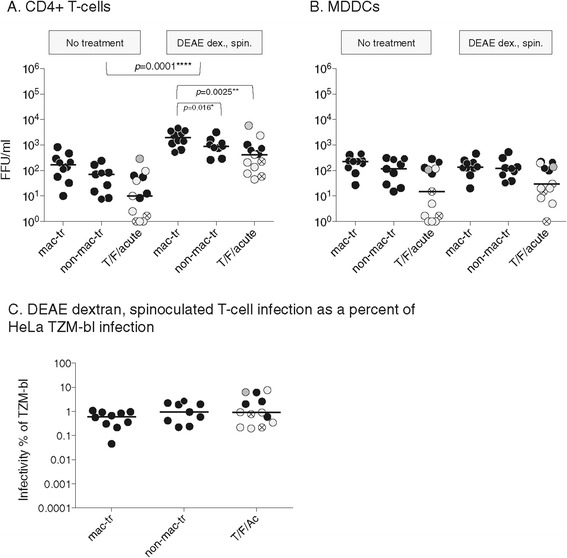
Results: Highly mac-tropic Envs mediated highest infectivity for primary T-cells, Jurkat/CCR5 cells, myeloid dendritic cells, macrophages, and HeLa TZM-bl cells, although this was most dramatic on macrophages. Infection of primary T-cells mediated by all Envs was low. However, infection of T-cells was greatly enhanced by increasing virus attachment with DEAE dextran and spinoculation, which enhanced the three Env+ virus groups to similar extents. Dendritic cell capture of viruses and trans-infection also greatly enhanced infection of primary T-cells. In trans-infection assays, non-mac-tropic R5 Envs were preferentially enhanced and those from late disease mediated levels of T-cell infection that were equivalent to those mediated by mac-tropic Envs.

Conclusions: Our results demonstrate that T/F, early or late disease non-mac-tropic R5 Envs do not preferentially mediate infection of primary CD4+ T-cells compared to highly mac-tropic Envs from brain tissue. We conclude that non-macrophage-tropism of HIV-1 R5 Envs in vitro is determined predominantly by a reduced capacity to target myeloid cells via low CD4 rather than a specific adaptation for T-cells entry that precludes macrophage infection.


Figures

Figure 1
Figure 1
Env+ pseudovirion infection of HeLa TZM-bl and macrophages. (A) Macrophage-tropic R5 Envs mediate 100-1000-fold higher infection of macrophages compared to T/F/acute and late disease non-mac-tropic R5 Envs. (B) Several late disease non-mac-tropic Envs and most T/F/acute Envs mediated lower infection of HeLa TZM-bl cells compared to macrophage tropic Envs. (C) Plotting macrophage infectivity titers as a percent of TZM-bl titers suggests that T/F/acute Envs are not as non-mac-tropic as late disease non-mac-tropic Envs. Significant differences were evaluated using Mann Whitney or Wilcoxon matched pair tests as described in Materials and Methods. Pseudoviruses carrying clade B Envs are represented by black symbols, those with clade C Envs by light gray symbols, clade A by hatched symbols and CRF A/C by dark gray. Median values are marked.
Figure 2
Figure 2
Infection of Jurkat/CCR5, primary CD4+ T-cells and primary MDDCs by pseudovirions carrying mac-tropic and non-mac-tropic R5 envelopes. Env+ GFP-reporter pseudovirus infection of Jurkat/CCR5 (A), primary CD4+ T-cells (B) and LPS matured MDDCs (C). Left panels shows FFU measured as GFP+ cells per ml of input virus. Right panels show infectivity as a percent of TZM-bl infectivity. Significant differences were evaluated using Mann Whitney or Wilcoxon matched pair tests as described in Methods. Only the p values that were significant are shown. Symbol colour designations are the same as described in Figure 1. Please also refer to Additional file 1: Figure S3, where FFU/ml for each Env+ pseudovirus is presented in bar graphs with standard deviation bars shown, while the column scatter plot of infectivity as a percent of TZM-bl infectivity is presented with each point labeled for Env used. Median values are marked.
Figure 3
Figure 3
DEAE dextran and spinoculation enhance infection for CD4+ T-cells but not for MDDCs. Infection in the presence and absence of DEAE dextran and spinoculation on CD4+ T-cells (A) and MDDCs (B). DEAE dextran and spinoculation enhance non-mac-tropic Envs and mac-tropic Envs to similar extents (C). Significant differences were evaluated using Mann Whitney or Wilcoxon matched pair tests as described in Materials and Methods. Figure 3 experiments were done in conjunction with the direct infections shown in Figure 2. Figure 3 data for infection without DEAE dextran and spinoculation is therefore the same as that shown in Figure 2. Only the p values relevant for DEAE dextran and spinoculation that show significance are shown. Symbol colour designations are the same as described in Figure 1. Please also refer to Additional file 1: Figure S4, where FFU/ml for each Env+ pseudovirus is presented in a bar graph with standard deviation bars shown, while the column scatter plot of infectivity as a percent of TZM-bl infectivity is presented with each point labeled for Env used. Median values are marked.
Figure 4
Figure 4
MDDC mediated trans-infection of CD4+ T-cells by Env+ pseudovirions. (A) Trans-infection of CD4+ T-cells following virion capture by MDDCs is substantially more efficient than cell free infection of T-cells or MDDCs alone. T/F/acute and late disease non-mac- and mac-tropic R5 Env+ pseudovirions are all enhanced. Note that for late disease non-mac-tropic and mac-tropic R5 Envs, trans-infectivity titers are not significantly different. Infectivities are shown as the number of FFU/ml (GFP+ cells per ml) of input virus. (B) Infectivity as a percent of infection for TZM-bl cells suggests that trans-infection preferentially enhances non-mac-tropic R5 Env+ pseudovirions, although differences are not significant. Symbol colour designations are the same as described in Figure 1. Please also refer to Additional file 1: Figure S5, where FFU/ml for each Env+ pseudovirus is presented in a bar graph with standard deviation bars shown, while the column scatter plot of infectivity as a percent of TZM-bl infectivity is presented with each point labeled for Env used. Median values are marked.

References

    1. Berger EA, Doms RW, Fenyo E-M, Korber BTM, Littman DR, Moore JP, et al. A new classification for HIV-1. Nature. 1998;391:240. doi: 10.1038/34571. - DOI - PubMed
    1. Peters PJ, Bhattacharya J, Hibbitts S, Dittmar MT, Simmons G, Bell J, et al. Biological analysis of human immunodeficiency virus type 1 R5 envelopes amplified from brain and lymph node tissues of AIDS patients with neuropathology reveals two distinct tropism phenotypes and identifies envelopes in the brain that confer an enhanced tropism and fusigenicity for macrophages. J Virol. 2004;78:6915–26. doi: 10.1128/JVI.78.13.6915-6926.2004. - DOI - PMC - PubMed
    1. Peters PJ, Sullivan WM, Duenas-Decamp MJ, Bhattacharya J, Ankghuambom C, Brown R, et al. Non-macrophage-tropic human immunodeficiency virus type 1 R5 envelopes predominate in blood, lymph nodes, and semen: implications for transmission and pathogenesis. J Virol. 2006;80:6324–32. doi: 10.1128/JVI.02328-05. - DOI - PMC - PubMed
    1. Koyanagi Y, Miles S, Mitsuyasu RT, Merrill JE, Vinters HV, Chen IS. Dual infection of the central nervous system by AIDS viruses with distinct cellular tropisms. Science. 1987;236:819–22. doi: 10.1126/science.3646751. - DOI - PubMed
    1. Dunfee R, Thomas ER, Gorry PR, Wang J, Ancuta P, Gabuzda D. Mechanisms of HIV-1 neurotropism. Curr HIV Res. 2006;4:267–78. doi: 10.2174/157016206777709500. - DOI - PubMed

Publication types

Substances