

JAK kinase inhibition abrogates STAT3 activation and head and neck squamous cell carcinoma tumor growth
- PMID: 25810010
- PMCID: PMC4372647
- DOI: 10.1016/j.neo.2015.01.003
JAK kinase inhibition abrogates STAT3 activation and head and neck squamous cell carcinoma tumor growth
Abstract
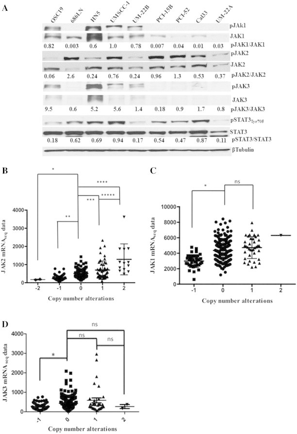
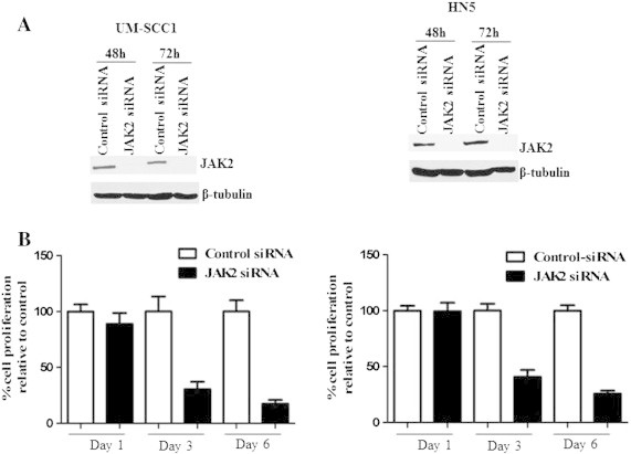
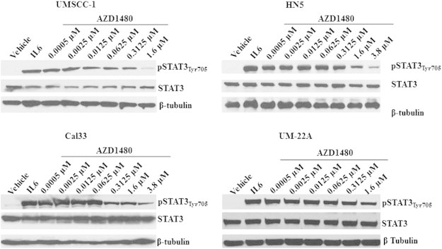
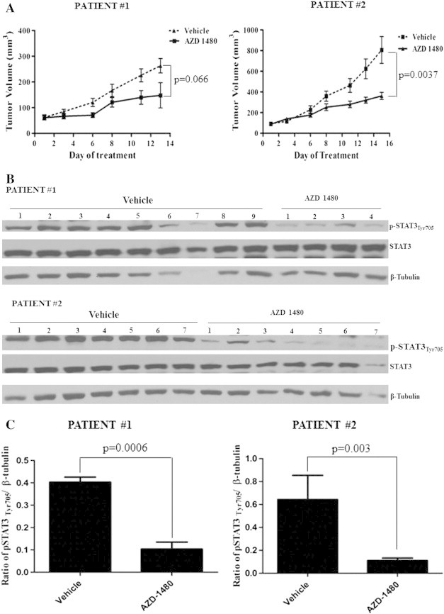
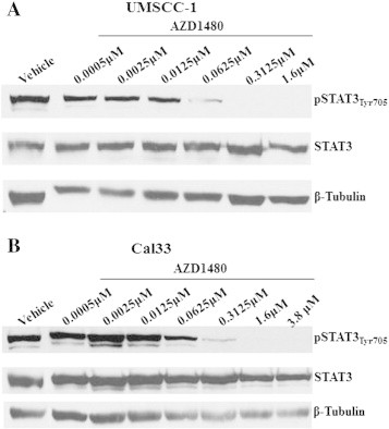
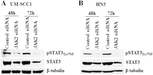
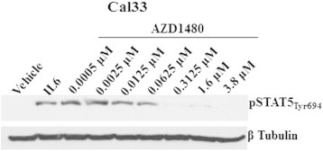
Aberrant activation of the Janus kinase (JAK)/signal transducer and activator of transcription (STAT) 3 has been implicated in cell proliferation and survival of many cancers including head and neck squamous cell carcinoma (HNSCC). AZD1480, an orally active pharmacologic inhibitor of JAK1/JAK2, has been tested in several cancer models. In the present study, the in vitro and in vivo effects of AZD1480 were evaluated in HNSCC preclinical models to test the potential use of JAK kinase inhibition for HNSCC therapy. AZD1480 treatment decreased HNSCC proliferation in HNSCC cell lines with half maximal effective concentration (EC50) values ranging from 0.9 to 4 μM in conjunction with reduction of pSTAT3(Tyr705) expression. In vivo antitumor efficacy of AZD1480 was demonstrated in patient-derived xenograft (PDX) models derived from two independent HNSCC tumors. Oral administration of AZD1480 reduced tumor growth in conjunction with decreased pSTAT3(Tyr705) expression that was observed in both PDX models. These findings suggest that the JAK1/2 inhibitors abrogate STAT3 signaling and may be effective in HNSCC treatment approaches.
Copyright © 2015 The Authors. Published by Elsevier Inc. All rights reserved.
Figures
Similar articles
-
The STAT3 pathway as a therapeutic target in head and neck cancer: Barriers and innovations.Oral Oncol. 2016 May;56:84-92. doi: 10.1016/j.oraloncology.2015.11.022. Epub 2015 Dec 28. Oral Oncol. 2016. PMID: 26733183 Free PMC article. Review.
-
p53-targeted lincRNA-p21 acts as a tumor suppressor by inhibiting JAK2/STAT3 signaling pathways in head and neck squamous cell carcinoma.Mol Cancer. 2019 Mar 11;18(1):38. doi: 10.1186/s12943-019-0993-3. Mol Cancer. 2019. PMID: 30857539 Free PMC article.
-
Inhibition of STAT3 with orally active JAK inhibitor, AZD1480, decreases tumor growth in Neuroblastoma and Pediatric Sarcomas In vitro and In vivo.Oncotarget. 2013 Mar;4(3):433-45. doi: 10.18632/oncotarget.930. Oncotarget. 2013. PMID: 23531921 Free PMC article.
-
Mutations of the LIM protein AJUBA mediate sensitivity of head and neck squamous cell carcinoma to treatment with cell-cycle inhibitors.Cancer Lett. 2017 Apr 28;392:71-82. doi: 10.1016/j.canlet.2017.01.024. Epub 2017 Jan 23. Cancer Lett. 2017. PMID: 28126323 Free PMC article.
-
Defining the role of the JAK-STAT pathway in head and neck and thoracic malignancies: implications for future therapeutic approaches.Drug Resist Updat. 2010 Jun;13(3):67-78. doi: 10.1016/j.drup.2010.04.001. Epub 2010 May 14. Drug Resist Updat. 2010. PMID: 20471303 Review.
Cited by
-
The STAT3 pathway as a therapeutic target in head and neck cancer: Barriers and innovations.Oral Oncol. 2016 May;56:84-92. doi: 10.1016/j.oraloncology.2015.11.022. Epub 2015 Dec 28. Oral Oncol. 2016. PMID: 26733183 Free PMC article. Review.
-
Malignancies in Patients with Celiac Disease: Diagnostic Challenges and Molecular Advances.Genes (Basel). 2023 Jan 31;14(2):376. doi: 10.3390/genes14020376. Genes (Basel). 2023. PMID: 36833303 Free PMC article. Review.
-
Toward the use of precision medicine for the treatment of head and neck squamous cell carcinoma.Oncotarget. 2017 Jan 10;8(2):2141-2152. doi: 10.18632/oncotarget.13798. Oncotarget. 2017. PMID: 27924064 Free PMC article. Review.
-
Head and neck cancer: pathogenesis and targeted therapy.MedComm (2020). 2024 Aug 21;5(9):e702. doi: 10.1002/mco2.702. eCollection 2024 Sep. MedComm (2020). 2024. PMID: 39170944 Free PMC article. Review.
-
Targeting Upstream Kinases of STAT3 in Human Medulloblastoma Cells.Curr Cancer Drug Targets. 2019;19(7):571-582. doi: 10.2174/1568009618666181016165604. Curr Cancer Drug Targets. 2019. PMID: 30332965 Free PMC article.
References
-
- Shuai K, Liu B. Regulation of JAK-STAT signalling in the immune system. Nat Rev Immunol. 2003;3:900–911. - PubMed
-
- Kupferman ME, Jayakumar A, Zhou G, Xie T, Dakak-Yazici Y, Zhao M, Ju J, Mandal M, Jasser S, Madden T. Therapeutic suppression of constitutive and inducible JAK\STAT activation in head and neck squamous cell carcinoma. J Exp Ther Oncol. 2009;8:117–127. - PubMed
-
- Seidel HM, Lamb P, Rosen J. Pharmaceutical intervention in the JAK/STAT signaling pathway. Oncogene. 2000;19:2645–2656. - PubMed
Publication types
MeSH terms
Substances
Grants and funding
LinkOut - more resources
Full Text Sources
Other Literature Sources
Medical
Research Materials
Miscellaneous
