Review
. 2015 Aug;26(8):1765-76.
doi: 10.1681/ASN.2015010006. Epub 2015 Mar 25.

Failed Tubule Recovery, AKI-CKD Transition, and Kidney Disease Progression

Affiliations
Review

Failed Tubule Recovery, AKI-CKD Transition, and Kidney Disease Progression

Manjeri A Venkatachalam et al. J Am Soc Nephrol. 2015 Aug.

Abstract

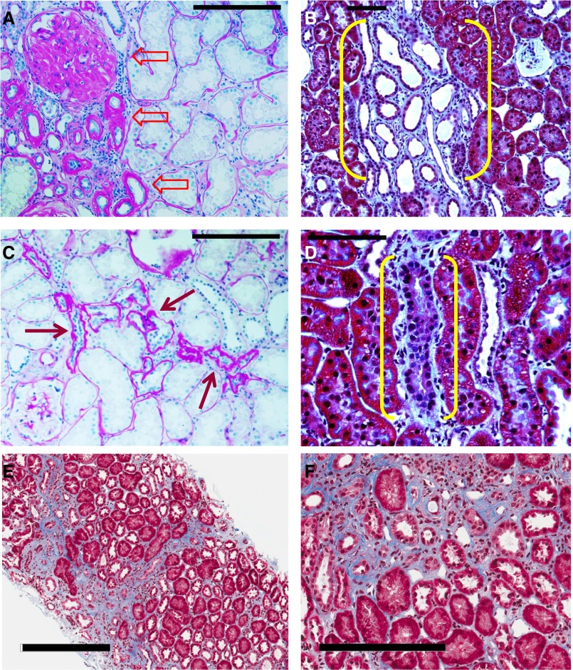
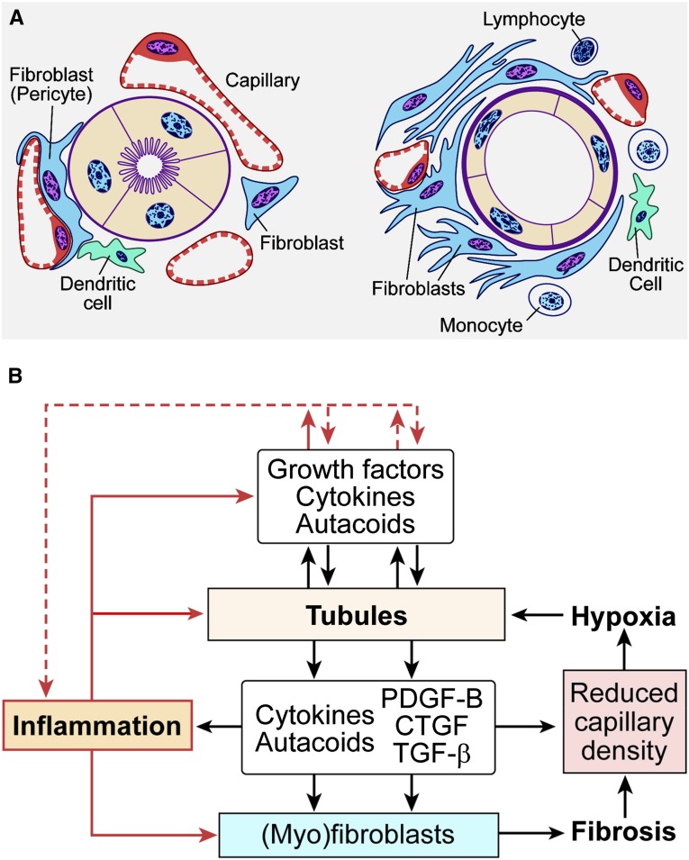
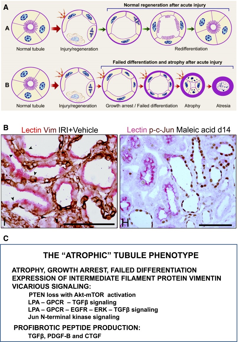
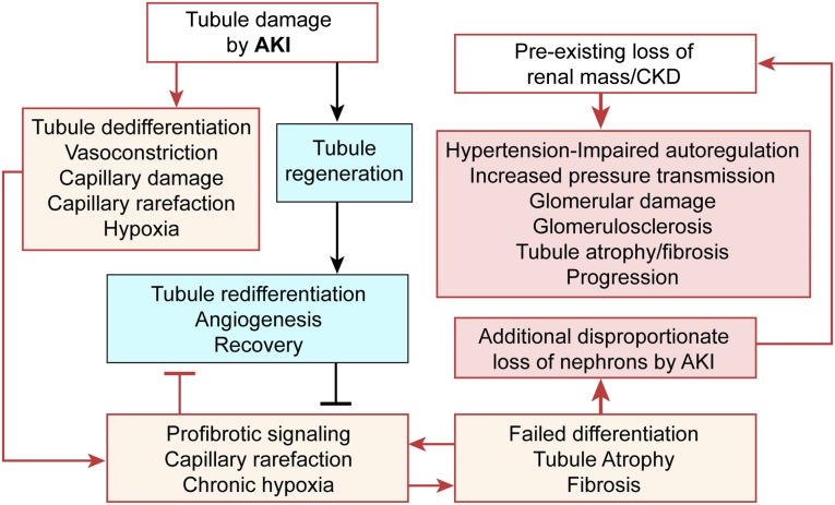
The transition of AKI to CKD has major clinical significance. As reviewed here, recent studies show that a subpopulation of dedifferentiated, proliferating tubules recovering from AKI undergo pathologic growth arrest, fail to redifferentiate, and become atrophic. These abnormal tubules exhibit persistent, unregulated, and progressively increasing profibrotic signaling along multiple pathways. Paracrine products derived therefrom perturb normal interactions between peritubular capillary endothelium and pericyte-like fibroblasts, leading to myofibroblast transformation, proliferation, and fibrosis as well as capillary disintegration and rarefaction. Although signals from injured endothelium and inflammatory/immune cells also contribute, tubule injury alone is sufficient to produce the interstitial pathology required for fibrosis. Localized hypoxia produced by microvascular pathology may also prevent tubule recovery. However, fibrosis is not intrinsically progressive, and microvascular pathology develops strictly around damaged tubules; thus, additional deterioration of kidney structure after the transition of AKI to CKD requires new acute injury or other mechanisms of progression. Indeed, experiments using an acute-on-chronic injury model suggest that additional loss of parenchyma caused by failed repair of AKI in kidneys with prior renal mass reduction triggers hemodynamically mediated processes that damage glomeruli to cause progression. Continued investigation of these pathologic mechanisms should reveal options for preventing renal disease progression after AKI.

Keywords: CKD; acute renal failure; fibrosis; hypertension; hypoxia; tubular epithelium.


Figures

Figure 1.
Figure 1.
Injured kidney tissue heals by fibrosis that does not extend to involve previously nondiseased parenchyma. (A and C) Periodic acid–Schiff staining of the kidney from an autopsy of a patient with human FSGS. (A) Advanced scar containing sclerotic glomerulus, atrophic tubules with greatly thickened basement membranes, and interstitial fibrosis is sharply demarcated from completely normal parenchyma (block arrows with red outlines). There is no indication of fibrosis spreading from the scar into the adjacent interstitium. (C) Small scar from the same kidney showing a few atrophic tubules with thick basement membranes and expanded interstitium adjacent to the atrophic tubules (red arrows) near a glomerulus with mild mesangial expansion. The histopathology is one of resolved injury to tubules with development of a shrunken scar in relationship to an atrophic nephron with no suggestion that the lesion is invasive or expansive in its nature. (B and D) Kidney of rat 14 days after AKI was induced by proximal tubule selective toxin maleic acid stained with Masson’s Trichrome. (B) Low-power micrograph showing a localized lesion containing undifferentiated atrophic tubules surrounded by a florid early fibrotic response (yellow brackets). The lesion is sharply demarcated from the adjacent well differentiated proximal tubules that had either recovered normally after AKI or had not been injured by the poison. (D) High-power micrograph showing a single profile of an atrophic tubule with surrounding fibroblastic response (yellow brackets). Adjacent proximal tubules are well differentiated. The interstitium between them is either normal or minimally expanded. Interstitial fibroblastic responses that occur after AKI resolve and regress as tubules recover and redifferentiate or persist and undergo scarring if tubules fail to redifferentiate and become atrophic. (E and F) Kidney biopsy from a patient 10 months after post-transplant AKI with delayed graft function stained with Masson’s Trichrome (provided by Robert B. Colvin, Massachusetts General Hospital and Harvard Medical School, Boston, MA). (E) Low-power micrograph showing shrunken mature scars separated by healthy parenchyma with minimal or no increase of interstitial connective tissue. (F) High-power micrograph with sharply demarcated boundary between scar and healthy tubules. One healthy well differentiated proximal tubule remains within the scar, suggesting that it had not been injured during the AKI episode 10 months earlier or had recovered normal structure during regeneration and repair after injury. Scale bars, 100 µm in A–D; 300 µm in E; 200 µm in F.
Figure 2.
Figure 2.
Pathologic events in tubules and interstitium interact to produce tubulointerstitial fibrosis. (A) Schematic diagram of (left panel) normal tubule-interstitium and (right panel) early tubulointerstitial fibrosis. Resident fibroblasts in the interstitium may or may not have intimate relationships to peritubular capillaries and basement membranes of tubules. The former type has also been termed pericyte. After injury, this type of fibroblast/pericyte detaches from capillaries, initiating pathologic events that cause capillary disintegration and rarefaction as well as myofibroblasts transformation and proliferation. This process is aided and abetted by inflammatory cells, chiefly monocytes, and resident immune cells, including dendritic cells. (B) Schematic diagram illustrating vicious cycle feedback interactions between tubule pathology and interstitial pathology that potentiate tubule atrophy. Modified from reference , with permission.
Figure 3.
Figure 3.
Failed differentiation of proximal tubules regenerating after AKI leads to development of the atrophic abnormally signaling profibrotic tubule phenotype. (A) Schematic diagram illustrating (upper panel) the normal pathway of proximal tubule cell dedifferentiation and proliferation followed by redifferentiation and recovery of normal structure after AKI and (lower panel) the abnormal pathway of failure to redifferentiate after early dedifferentiation that leads to tubule atrophy after AKI. (B, left panel) Immunohistochemistry and PHA lectin affinity cytochemistry of atrophic and normal proximal tubule cells 14 days after IRI in rats. Mosaic tubules showing well differentiated proximal tubule cells with brush border–bound PHA (lectin) staining pink in close juxtaposition with atrophic epithelium without brush border that stains brown for the expression of vimentin, an intermediate filament protein that is not present in differentiated proximal tubule cells but is rapidly expressed after dedifferentiation during regeneration and retained after atrophy occurs. (B, right panel) Immunohistochemistry and lectin cytochemistry of proximal tubules 14 days after AKI induced by proximal tubule selective toxin maleic acid. Well differentiated proximal tubule profiles and well differentiated proximal tubule cells in a mosaic tubule (center) exhibit pink staining for brush border–bound PHA lectin but no nuclear staining for phospho-c-Jun, whereas atrophic tubule profiles and atrophic cells in mosaic tubule (center) show nuclear staining for phospho-c-Jun, indicating the activation of the JNK-MAPK signaling pathway. (C) The diverse abnormalities exhibited by atrophic tubules are listed. These several alterations take place in vimentin–expressing atrophic tubules illustrated in B, left panel. Scale bars, 100 µm. A is modified from reference , with permission. B is modified from reference , with permission. GPCR, G-protein coupled receptor; JNK-MAPK, Jun N-terminal kinase-mitogen activated protein kinase; LPA, lysophosphatidic acid; PHA, phytohemagglutinin; PTEN, phosphatase and tensin homolog.
Figure 4.
Figure 4.
Failed tubule differentiation and RMR after AKI lead to hemodynamic abnormalities that cause progression. Schematic diagram illustrating the effects of AKI that lead to tubulointerstitial fibrosis, the RMR that retards recovery of tubules regenerating after AKI, and the resulting disproportionate further reduction of renal mass that triggers hemodynamic mechanisms of renal disease progression.

References

    1. Hsu CY: Yes, AKI truly leads to CKD. J Am Soc Nephrol 23: 967–969, 2012 - PubMed
    1. Okusa MD, Chertow GM, Portilla D, Acute Kidney Injury Advisory Group of the American Society of Nephrology : The nexus of acute kidney injury, chronic kidney disease, and World Kidney Day 2009. Clin J Am Soc Nephrol 4: 520–522, 2009 - PMC - PubMed
    1. Chawla LS, Eggers PW, Star RA, Kimmel PL: Acute kidney injury and chronic kidney disease as interconnected syndromes. N Engl J Med 371: 58–66, 2014 - PMC - PubMed
    1. Venkatachalam MA, Griffin KA, Lan R, Geng H, Saikumar P, Bidani AK: Acute kidney injury: A springboard for progression in chronic kidney disease. Am J Physiol Renal Physiol 298: F1078–F1094, 2010 - PMC - PubMed
    1. Bydash JR, Ishani A: Acute kidney injury and chronic kidney disease: A work in progress. Clin J Am Soc Nephrol 6: 2555–2557, 2011 - PubMed

MeSH terms