

Pandemic Swine H1N1 Influenza Viruses with Almost Undetectable Neuraminidase Activity Are Not Transmitted via Aerosols in Ferrets and Are Inhibited by Human Mucus but Not Swine Mucus
- PMID: 25810540
- PMCID: PMC4442420
- DOI: 10.1128/JVI.02537-14
Pandemic Swine H1N1 Influenza Viruses with Almost Undetectable Neuraminidase Activity Are Not Transmitted via Aerosols in Ferrets and Are Inhibited by Human Mucus but Not Swine Mucus
Abstract
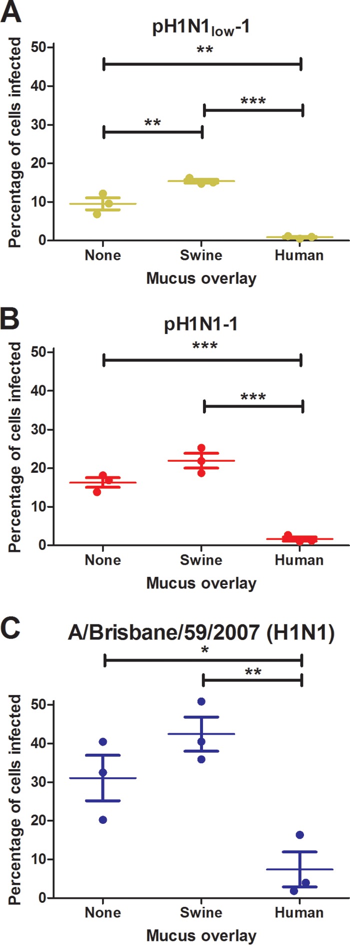
A balance between the functions of the influenza virus surface proteins hemagglutinin (HA) and neuraminidase (NA) is thought to be important for the transmission of viruses between humans. Here we describe two pandemic H1N1 viruses, A/swine/Virginia/1814-1/2012 and A/swine/Virginia/1814-2/2012 (pH1N1low-1 and -2, respectively), that were isolated from swine symptomatic for influenza. The enzymatic activity of the NA of these viruses was almost undetectable, while the HA binding affinity for α2,6 sialic acids was greater than that of the highly homologous pH1N1 viruses A/swine/Pennsylvania/2436/2012 and A/swine/Minnesota/2499/2012 (pH1N1-1 and -2), which exhibited better-balanced HA and NA activities. The in vitro growth kinetics of pH1N1low and pH1N1 viruses were similar, but aerosol transmission of pH1N1low-1 was abrogated and transmission via direct contact in ferrets was significantly impaired compared to pH1N1-1, which transmitted by direct and aerosol contact. In normal human bronchial epithelial cells, pH1N1low-1 was significantly inhibited by mucus but pH1N1-1 was not. In Madin-Darby canine kidney cell cultures overlaid with human or swine mucus, human mucus inhibited pH1N1low-1 but swine mucus did not. These data show that the interaction between viruses and mucus may be an important factor in viral transmissibility and could be a barrier for interspecies transmission between humans and swine for influenza viruses.
Importance: A balance between the functions of the influenza virus surface proteins hemagglutinin (HA) and neuraminidase (NA) is thought to be important for transmission of viruses from swine to humans. Here we show that a swine virus with extremely functionally mismatched HA and NAs (pH1N1low-1) cannot transmit via aerosol in ferrets, while another highly homologous virus with HA and NAs that are better matched functionally (pH1N1-1) can transmit via aerosol. These viruses show similar growth kinetics in Madin-Darby canine kidney (MDCK) cells, but pH1N1low-1 is significantly inhibited by mucus in normal human bronchial epithelial cells whereas pH1N1-1 is not. Further, human mucus could inhibit these viruses, but swine mucus could not. These data show that the interaction between viruses and mucus may be an important factor in viral transmissibility and could be a species barrier between humans and swine for influenza viruses.
Copyright © 2015, American Society for Microbiology. All Rights Reserved.
Figures
References
-
- Yen HL, Liang CH, Wu CY, Forrest HL, Ferguson A, Choy KT, Jones J, Wong DD, Cheung PP, Hsu CH, Li OT, Yuen KM, Chan RW, Poon LL, Chan MC, Nicholls JM, Krauss S, Wong CH, Guan Y, Webster RG, Webby RJ, Peiris M. 2011. Hemagglutinin-neuraminidase balance confers respiratory-droplet transmissibility of the pandemic H1N1 influenza virus in ferrets. Proc Natl Acad Sci U S A 108:14264–14269. doi:10.1073/pnas.1111000108. - DOI - PMC - PubMed
Publication types
MeSH terms
Substances
Grants and funding
LinkOut - more resources
Full Text Sources
