

Infra-orbital nerve schwannoma: Report and review
- PMID: 25810682
- PMCID: PMC4367058
- DOI: 10.4103/0976-9668.149229
Infra-orbital nerve schwannoma: Report and review
Abstract
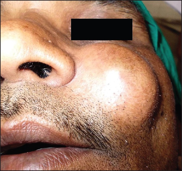
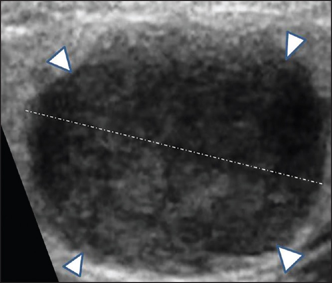
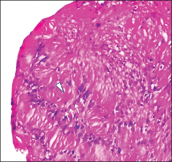
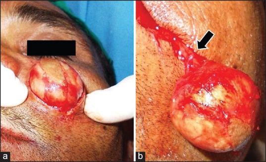
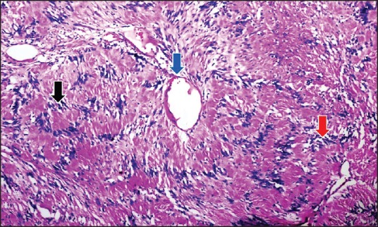
Extra-cranial schwannomas although common in head and neck region are very rarely seen originating from the infra-orbital nerve. We report a case of schwannoma arising from infra-orbital nerve in a 40-year-old male patient. The case presented as an isolated, asymptomatic, slow growing sub-cutaneous nodular swelling over left side of mid-face. On ultrasonography, a localized lesion within the sub-cutaneous tissue of cheek was observed, without involvement of orbital, maxillary sinus or underlying bone. Aspiration biopsy of the lesion showed spindle shaped cells predominantly arranged in Antoni A pattern around verocay bodies, with less organized Antoni B tissue in few places. Diagnosis of schwannoma, probably arising from terminal branch of infra-orbital nerve was established. The tumor was approached through skin incision. At the time of exploration, the lesion was found to emanate from the nerve trunk of peripheral branch of infra-orbital nerve, which was dissected and preserved. We correlate our experience with previously reported cases of infra-orbital nerve schwannoma.
Keywords: Infra-orbital nerve; mid-face; schwannoma; swelling.
Conflict of interest statement
Figures
References
-
- Marx RE, Stern D. United States Quintessence Publishing Co. Inc. 2nd ed 2003. Oral and Maxillofacial Pathology: A Rationale for Diagnosis and Treatment.
-
- Samet A, Podoshin L, Fradis M, Simon J, Lazarov N, Boss H. Unusual sites of schwannoma in the head and neck. J Laryngol Otol. 1985;99:523–8. - PubMed
-
- Choi BH, Park SW, Son JH, Cho YC, Sung IY, Byun KJ, et al. Schwannoma in the maxillary sinus and buccal space: Case report. J Korean Assoc Oral Maxillofac Surg. 2009;35:494–8.
-
- Yoon E, Rhee SC. Solitary trigeminal schwannoma of paranasal region. Int J Pediatric Otorhinolaryngol Extra 01. 2007;2:120–4.
-
- Leu YS, Chang KC. Extracranial head and neck schwannomas: A review of 8 years experience. Acta Otolaryngol. 2002;122:435–7. - PubMed
Publication types
LinkOut - more resources
Full Text Sources
Other Literature Sources