. 2015 Mar 3;49(1):41-9.
doi: 10.2478/raon-2014-0045. eCollection 2015 Mar.

Mild hyperthermia influence on Herceptin(®) properties

Affiliations

Mild hyperthermia influence on Herceptin(®) properties

Jean-Michel Escoffre et al. Radiol Oncol. .

Abstract

Background: Mild hyperthermia (mHT) increases the tumor perfusion and vascular permeability, and reduces the interstitial fluid pressure, resulting in better intra-tumoral bioavailability of low molecular weight drugs. This approach is potentially also attractive for delivery of therapeutic macromolecules, such as antibodies. Here, we investigated the effects of mHT on the stability, immunological and pharmacological properties of Herceptin(®), a clinically approved antibody, targeting the human epidermal growth factor receptor 2 (HER-2) overexpressed in breast cancer.

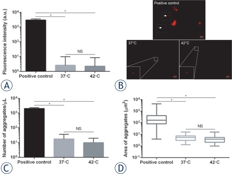
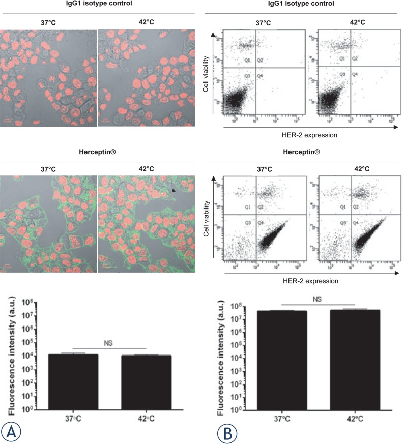
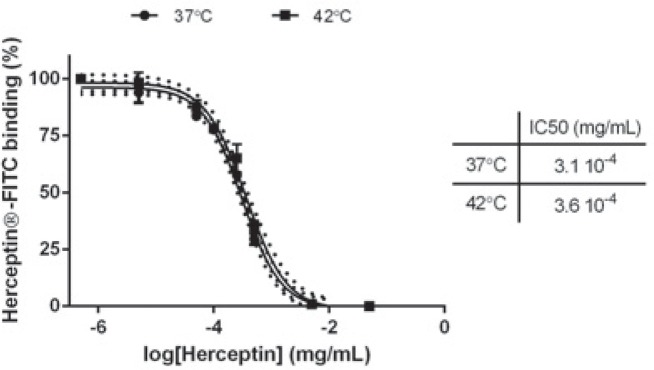
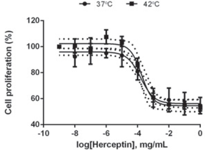
Results: Herceptin(®) was heated to 37°C (control) and 42°C (mHT) for 1 hour. Formation of Herceptin(®) aggregates was measured using Nile Red assay. mHT did not result in additional Herceptin(®) aggregates compared to 37°C, showing the Herceptin(®) stability is unchanged. Immunological and pharmacological properties of Herceptin(®) were evaluated following mHT using HER-2 positive breast cancer cells (BT-474). Exposure of Herceptin(®) to mHT preserved recognition and binding affinity of Herceptin(®) to HER-2. Western-blot and cell proliferation assays on BT-474 cells showed that mHT left the inhibitory activities of Herceptin(®) unchanged.

Conclusions: The stability, and the immunological and pharmacological properties of Herceptin(®) are not negatively affected by mHT. Further in-vivo studies are required to evaluate the influence of mHT on intra-tumoral bioavailability and therapeutic effectiveness of Herceptin(®).

Keywords: Herceptin®; anticancer antibody; breast cancer; mild hyperthermia.


Figures

FIGURE 1.
FIGURE 1.
Herceptin® stability. Immediately after Nile Red addition (100 μM), 0.8 mg/mL antibody solution was incubated at 37°C (native antibody), 42°C (heated antibody) and 90°C (positive control) for 1 h. Spectrofluorimetry was used for measuring total fluorescence signal (A), whereas fluorescence images were taken for observing Herceptin® aggregates (B). Representative images of antibody aggregates for each experimental condition are shown. Concentration (C) and area (D) of antibody aggregates are reported. Data expressed as mean ± SD was calculated from five independent experiments. Statistical analysis was performed using the non-parametric Mann-Whitney test. Significance was defined as p < 0.05 (NS, non-significant; *p < 0.05 compared to the positive condition).
FIGURE 2.
FIGURE 2.
Recognition of HER-2 by Herceptin® following mHT. BT-474 cells were incubated with 10 μg/mL native or heated Herceptin®, or matched human isotype control IgG1. Subsequently, the cells were incubated with FITC-conjugated anti-human IgG1-Fc antibody and then analyzed by confocal microscopy (A) and flow cytometry (B). Representative images of BT-474 cells and dot-plots of each experimental condition are shown. Data expressed as mean ± SD was calculated from three independent experiments. Statistical analysis was performed using the non-parametric Mann-Whitney test. Significance was defined as p < 0.05 (NS, non-significant).
FIGURE 3.
FIGURE 3.
Binding affinity of Herceptin® to HER-2. BT-474 cells were first incubated with unconjugated Herceptin® (5 × 10⁻⁶ to 5 × 10⁻² mg/mL) and subsequently with FITC-Herceptin®. Fluorescence intensity on flow cytometry is plotted as a function of unlabeled Herceptin® concentration used for receptor saturation. Data expressed as mean ± SD calculated from three independent experiments and are fitted with Variable slope model (solid curve; confidence intervals, dotted curve) with a 95% confidence interval. Statistical analysis was performed using the non-parametric Mann-Whitney test. Significance was defined as p < 0.05 (NS, non-significant).
FIGURE 4.
FIGURE 4.
Degradation and Dephosphorylation of HER-2 receptors by Herceptin®. 4×10⁵ BT-474 cells were seeded in a 6-well plate and treated with Herceptin® (50 μg/mL) for 6 days. Immunoblots of protein lysates were analyzed for total and phosphorylated HER-2 receptors. Representative immunoblots of one experiment out of three independent experiments are shown. The number at the bottom of each lane indicates the relative fold change versus the control after normalization with the β-ACTIN signal. Data expressed as mean ± SD calculated from three independent experiments. Statistical analysis was performed using the non-parametric Mann-Whitney test. Significance was defined as p < 0.05 (NS, non-significant; *p < 0.05 compared to the positive condition).
FIGURE 5.
FIGURE 5.
Effect of Herceptin® on BT-474 cell proliferation. BT-474 cells were seeded in a 96-well plate at 15,000 cells per well and were treated with native or heated Herceptin® (1×10⁻⁸ - 5×10⁻² mg/mL) for 6 days. Cell proliferation assay was performed. The mean relative growth was expressed as a ratio of the mean control relative growth (vehicle-treated cells). Data expressed as mean ± SD calculated from four independent experiments and are fitted with Variable slope model (solid curve; confidence intervals, dotted curve) with a 95% confidence interval. Statistical analysis was performed using the non-parametric Mann-Whitney test. Significance was defined as p < 0.05 (NS, non-significant).

References

    1. Ferlay J, Steliarova-Foucher E, Lortet-Tieulent J, Rosso S, Coebergh JW, Comber H, et al. Cancer incidence and mortality patterns in Europe: estimates for 40 countries in 2012. Eur J Cancer. 2013;49:1374–403. - PubMed
    1. Arteaga CL, Sliwkowski MX, Osborne CK, Perez EA, Puglisi F, Gianni L. Treatment of HER2-positive breast cancer: current status and future perspectives. Nat Rev Clin Oncol. 2012;9:16–32. - PubMed
    1. Eroles P, Bosch A, Perez-Fidalgo JA, Lluch A. Molecular biology in breast cancer: intrinsic subtypes and signaling pathways. Cancer Treat Rev. 2012;38:698–707. - PubMed
    1. Yardley DA, Tripathy D, Brufsky AM, Rugo HS, Kaufman PA, Mayer M, et al. Long-term survivor characteristics in HER2-positive metastatic breast cancer from registHER. Br J Cancer. 2014;110:2756–64. - PMC - PubMed
    1. Hudis CA. Trastuzumab-mechanism of action and use in clinical practice. N Engl J Med. 2007;357:39–51. - PubMed