

Neuroprotective and antiapoptotic activity of lineage-negative bone marrow cells after intravitreal injection in a mouse model of acute retinal injury
- PMID: 25810725
- PMCID: PMC4354968
- DOI: 10.1155/2015/620364
Neuroprotective and antiapoptotic activity of lineage-negative bone marrow cells after intravitreal injection in a mouse model of acute retinal injury
Abstract
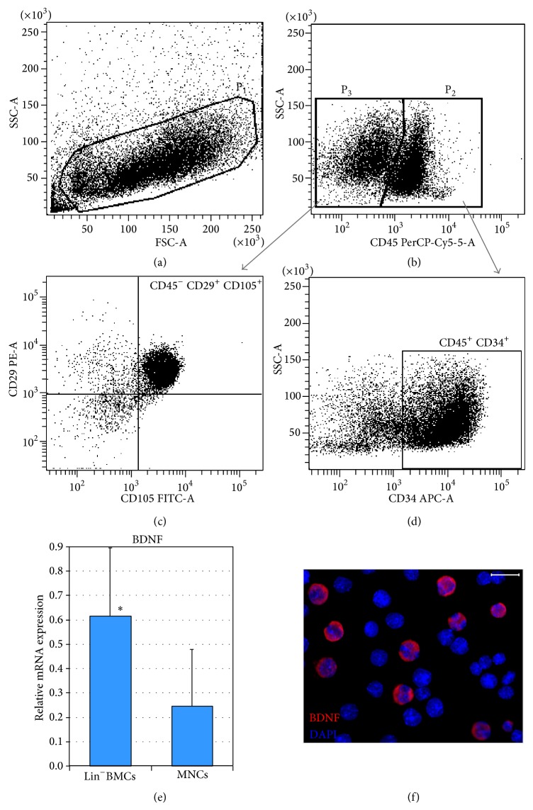
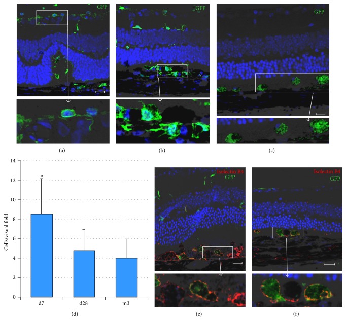
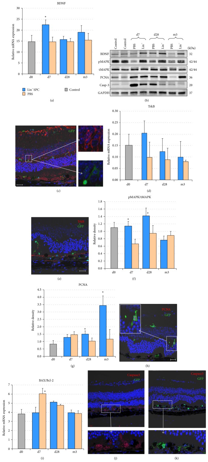
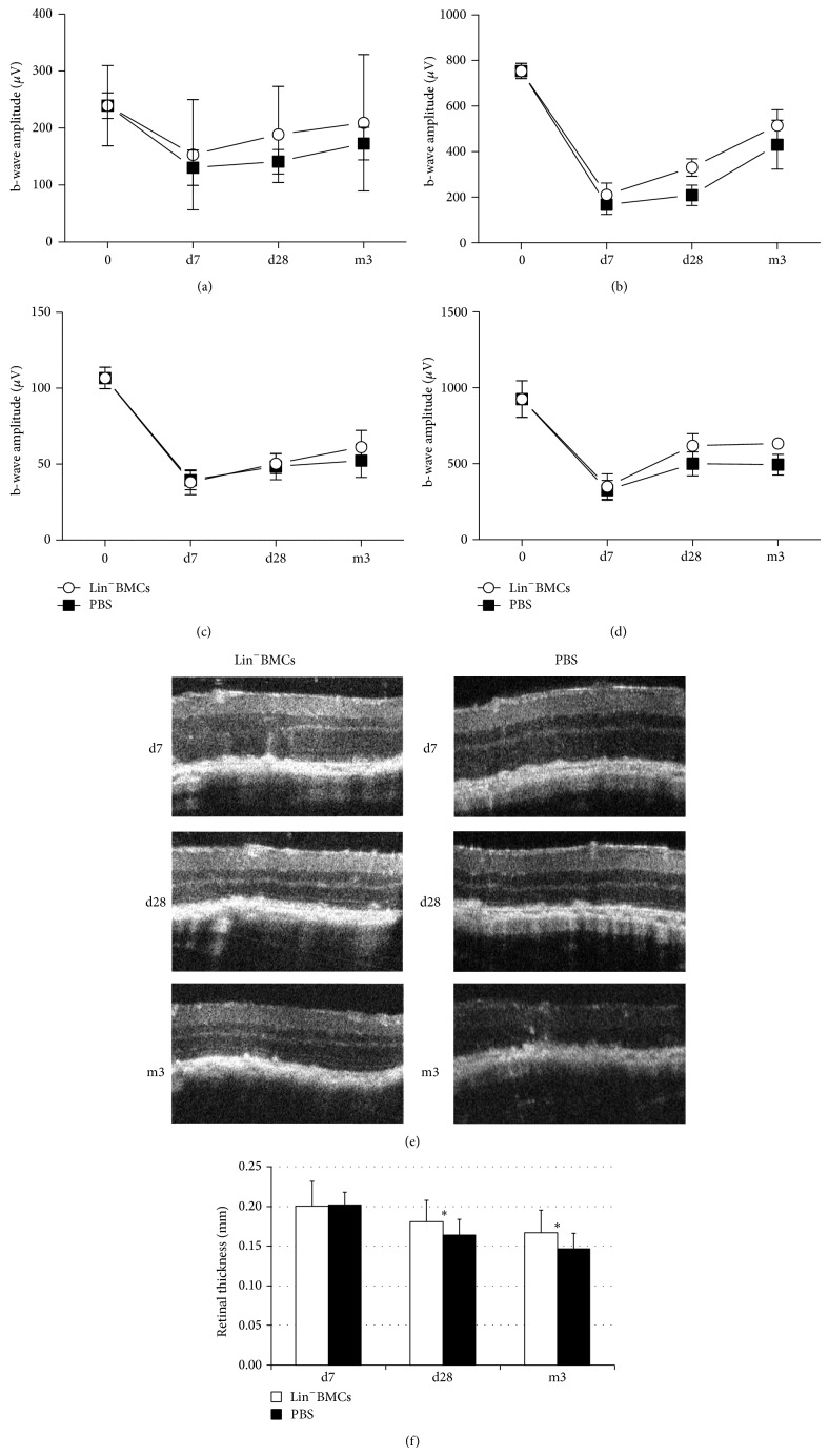
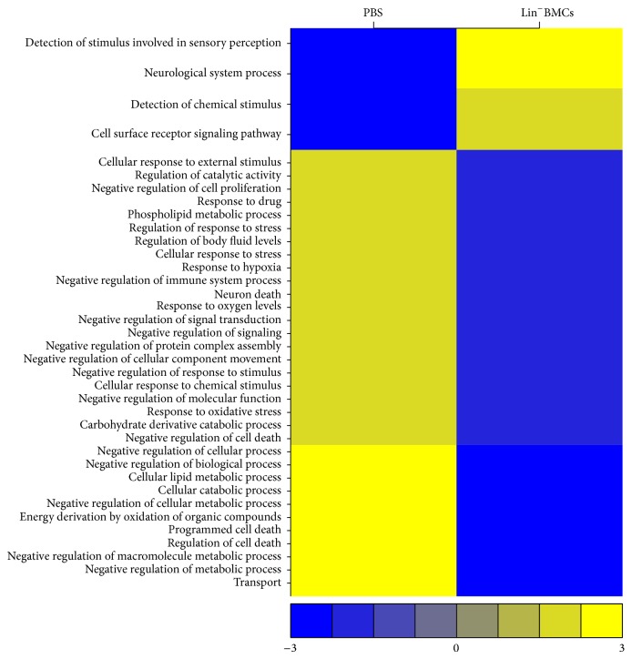
We investigated effects of bone marrow-derived, lineage-negative cell (Lin(-)BMC) transplantation in acute retinal injury. Lin(-)BMCs were intravitreally injected into murine eyes at 24 h after NaIO3-induced injury. Morphology, function, and expression of apoptosis-related genes, including brain-derived neurotrophic factor (BDNF) and its receptor, were assessed in retinas at 7 days, 28 days, and 3 months after transplantation. Moreover, global gene expression at day 7 was analyzed by RNA arrays. We observed that Lin(-)BMCs integrated into outer retinal layers improving morphological retinal structure and induced molecular changes such as downregulation of proapoptotic caspase-3 gene, a decrease in BAX/BCL-2 gene ratio, and significant elevation of BDNF expression. Furthermore, transplanted Lin(-)BMCs differentiated locally into cells with a macrophage-like phenotype. Finally, Lin(-)BMCs treatment was associated with generation of two distinct transcriptomic patterns. The first relates to downregulated genes associated with regulation of neuron cell death and apoptosis, response to oxidative stress/hypoxia and external stimuli, and negative regulation of cell proliferation. The second relates to upregulated genes associated with neurological system processes and sensory perception. Collectively, our data demonstrate that transplanted Lin(-)BMCs exert neuroprotective function against acute retinal injury and this effect may be associated with their antiapoptotic properties and ability to express neurotrophic factors.
Figures
Similar articles
-
Preclinical Evaluation of Long-Term Neuroprotective Effects of BDNF-Engineered Mesenchymal Stromal Cells as Intravitreal Therapy for Chronic Retinal Degeneration in Rd6 Mutant Mice.Int J Mol Sci. 2019 Feb 12;20(3):777. doi: 10.3390/ijms20030777. Int J Mol Sci. 2019. PMID: 30759764 Free PMC article.
-
Alteration of Neurotrophic Factors After Transplantation of Bone Marrow Derived Lin-ve Stem Cell in NMDA-Induced Mouse Model of Retinal Degeneration.J Cell Biochem. 2017 Jul;118(7):1699-1711. doi: 10.1002/jcb.25827. Epub 2017 Jan 11. J Cell Biochem. 2017. PMID: 27935095
-
Intramuscular transplantation and survival of freshly isolated bone marrow cells following skeletal muscle ischemia-reperfusion injury.J Trauma Acute Care Surg. 2013 Aug;75(2 Suppl 2):S142-9. doi: 10.1097/TA.0b013e31829ac1fa. J Trauma Acute Care Surg. 2013. PMID: 23883899
-
Transplantation of lineage-negative stem cells in pterygopalatine artery ligation induced retinal ischemia-reperfusion injury in mice.Mol Cell Biochem. 2017 May;429(1-2):123-136. doi: 10.1007/s11010-017-2941-0. Epub 2017 Feb 16. Mol Cell Biochem. 2017. PMID: 28210901
-
[Autologous bone marrow cell infusion therapy for liver cirrhosis--now and future].Rinsho Byori. 2011 Dec;59(12):1092-8. Rinsho Byori. 2011. PMID: 22338911 Review. Japanese.
Cited by
-
Preclinical Evaluation of Long-Term Neuroprotective Effects of BDNF-Engineered Mesenchymal Stromal Cells as Intravitreal Therapy for Chronic Retinal Degeneration in Rd6 Mutant Mice.Int J Mol Sci. 2019 Feb 12;20(3):777. doi: 10.3390/ijms20030777. Int J Mol Sci. 2019. PMID: 30759764 Free PMC article.
-
Electrostatic complex of neurotrophin 4 with dendrimer nanoparticles: controlled release of protein in vitro and in vivo.Int J Nanomedicine. 2019 Aug 1;14:6117-6131. doi: 10.2147/IJN.S210140. eCollection 2019. Int J Nanomedicine. 2019. PMID: 31534337 Free PMC article.
-
Therapeutic efficacy of neural stem cells originating from umbilical cord-derived mesenchymal stem cells in diabetic retinopathy.Sci Rep. 2017 Mar 24;7(1):408. doi: 10.1038/s41598-017-00298-2. Sci Rep. 2017. PMID: 28341839 Free PMC article.
-
Human Dental Pulp Stem Cells (DPSCs) Therapy in Rescuing Photoreceptors and Establishing a Sodium Iodate-Induced Retinal Degeneration Rat Model.Tissue Eng Regen Med. 2021 Feb;18(1):143-154. doi: 10.1007/s13770-020-00312-1. Epub 2021 Jan 7. Tissue Eng Regen Med. 2021. PMID: 33415670 Free PMC article.
-
Loss of Angiotensin-Converting Enzyme 2 Exacerbates Diabetic Retinopathy by Promoting Bone Marrow Dysfunction.Stem Cells. 2018 Sep;36(9):1430-1440. doi: 10.1002/stem.2848. Epub 2018 Jul 15. Stem Cells. 2018. PMID: 29761600 Free PMC article.
References
-
- Drela K., Siedlecka P., Sarnowska A., Domanska-Janik K. Human mesenchymal stem cells in the treatment of neurological diseases. Acta Neurobiologiae Experimentalis. 2013;73(1):38–56. - PubMed
LinkOut - more resources
Full Text Sources
Other Literature Sources
Research Materials
