. 2015 Apr;21(4):650-4.
doi: 10.3201/eid2104.141859.

Pathogenicity of 2 porcine deltacoronavirus strains in gnotobiotic pigs

Pathogenicity of 2 porcine deltacoronavirus strains in gnotobiotic pigs

Kwonil Jung et al. Emerg Infect Dis. 2015 Apr.

Abstract

To verify whether porcine deltacoronavirus infection induces disease, we inoculated gnotobiotic pigs with 2 virus strains (OH-FD22 and OH-FD100) identified by 2 specific reverse transcription PCRs. At 21-120 h postinoculation, pigs exhibited severe diarrhea, vomiting, fecal shedding of virus, and severe atrophic enteritis. These findings confirm that these 2 strains are enteropathogenic in pigs.

Keywords: PCR; PDCoV; coronavirus; gnotobiotic pigs; pathogenicity; pigs; porcine deltacoronavirus; strain OH-FD100; strain OH-FD22; strains; viruses.


Figures
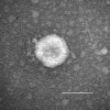
Figure 1
Figure 1
Porcine deltacoronavirus (OH-FD22) particle detected in intestinal contents from a gnotobiotic pig. The sample was negatively stained with 3% phosphotungstic acid. Scale bar indicates 100 nm.
Figure 2
Figure 2
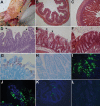
Intestinal changes in gnotobiotic pigs inoculated with porcine deltacoronavirus (PDCoV) strains OH-FD22 (panels A, B, E, G, I, and J) and OH-FD100 (panels D and K). A) Intestine of pig 3 at hour postinoculation (hpi) 72 (48–51 h after onset of clinical signs), showing thin and transparent intestinal walls (duodenum to colon) and accumulation of large amounts of yellow fluid in the intestinal lumen (arrows). B) Jejunum of pig 3 at hpi 72 (48–51 h after onset of clinical signs), showing acute diffuse, severe atrophic jejunitis (original magnification ×40). C) Jejunum of noninoculated pig 7, showing normal villous epithelium (original magnification ×80). D) Jejunum of pig 4 at hpi 96 (72–74 h after onset of clinical signs), showing acute diffuse, severe atrophic jejunitis with mild cytoplasmic vacuolation at the tips of villi (arrows) (original magnification ×200). E) Colon of pig 3 at hpi 72 (48–51 h after onset of clinical signs), showing mild cytoplasmic vacuolation of superficial epithelial cells (arrows) (original magnification ×200). F) Colon of noninoculated pig 7, showing normal colonic epithelium (original magnification ×200). G) Jejunum of pig 3 at hpi 72 (48–51 h after onset of clinical signs), showing that epithelial cells lining the atrophied villi are positive for PDCoV RNA (original magnification ×200). H) Jejunum of noninoculated pig 6, showing absence of PDCoV RNA-positive cells and background staining (original magnification ×200). I) Jejunum of pig 3 at hpi 72 (48–51 h after onset of clinical signs), showing large numbers of PDCoV antigen–positive cells in the epithelium of atrophied villi (original magnification ×200). J) Jejunum of pig 3 at hpi 72 (48–51 h after onset of clinical signs), showing localization of PDCoV antigens in cytoplasm of columnar epithelial cells (original magnification ×400). K) Cecum of pig 4 at hpi 96 (72–74 h after onset of clinical signs), showing a few PDCoV antigen–positive cells in the epithelium (original magnification ×200. L) Jejunum of noninoculated pig 6, showing absence of immunofluorescence-stained cells and background staining (original magnification ×200). Nuclei were stained with blue-fluorescent 4′, 6-diamidino-2-phenylindole dihydrochloride. Hematoxylin and eosin staining in panels B–F; in situ hybridization staining in panels G and H; immunofluorescence staining in panels I–L.

References

    1. Cima G. Viral disease affects U.S. pigs: porcine epidemic diarrhea found in at least 11 states. J Am Vet Med Assoc. 2013;243:30–1 . - PubMed
    1. Stevenson GW, Hoang H, Schwartz KJ, Burrough ER, Sun D, Madson D, et al. Emergence of porcine epidemic diarrhea virus in the United States: clinical signs, lesions, and viral genomic sequences. J Vet Diagn Invest. 2013;25:649–54. 10.1177/1040638713501675 - DOI - PubMed
    1. Marthaler D, Jiang Y, Collins J, Rossow K. Complete genome sequence of strain SDCV/USA/Illinois121/2014, a porcine deltacoronavirus from the United States. Genome Announc. 2014;2:e00218–14. - PMC - PubMed
    1. Wang L, Byrum B, Zhang Y. Detection and genetic characterization of deltacoronavirus in pigs, Ohio, USA, 2014. Emerg Infect Dis. 2014;20:1227–30. 10.3201/eid2007.140296 - DOI - PMC - PubMed
    1. Li G, Chen Q, Harmon KM, Yoon KJ, Schwartz KJ, Hoogland MJ, et al. Full-length genome sequence of porcine deltacoronavirus strain USA/IA/2014/8734. Genome Announc. 2014;2:e00278–14. - PMC - PubMed

Publication types