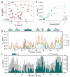
. 2015 Apr 28;7(16):7197-205.
doi: 10.1039/c5nr00539f.

Polyhydroxylated [60]fullerene binds specifically to functional recognition sites on a monomeric and a dimeric ubiquitin

Affiliations

Polyhydroxylated [60]fullerene binds specifically to functional recognition sites on a monomeric and a dimeric ubiquitin

Serena Zanzoni et al. Nanoscale. .

Abstract

The use of nanoparticles (NPs) in biomedical applications requires an in-depth understanding of the mechanisms by which NPs interact with biomolecules. NPs associating with proteins may interfere with protein-protein interactions and affect cellular communication pathways, however the impact of NPs on biomolecular recognition remains poorly characterized. In this respect, particularly relevant is the study of NP-induced functional perturbations of proteins implicated in the regulation of key biochemical pathways. Ubiquitin (Ub) is a prototypical protein post-translational modifier playing a central role in numerous essential biological processes. To contribute to the understanding of the interactions between this universally distributed biomacromolecule and NPs, we investigated the adsorption of polyhydroxylated [60]fullerene on monomeric Ub and on a minimal polyubiquitin chain in vitro at atomic resolution. Site-resolved chemical shift and intensity perturbations of Ub's NMR signals, together with (15)N spin relaxation rate changes, exchange saturation transfer effects, and fluorescence quenching data were consistent with the reversible formation of soluble aggregates incorporating fullerenol clusters. The specific interaction epitopes were identified, coincident with functional recognition sites in a monomeric and lysine48-linked dimeric Ub. Fullerenol appeared to target the open state of the dynamic structure of a dimeric Ub according to a conformational selection mechanism. Importantly, the protein-NP association prevented the enzyme-catalyzed synthesis of polyubiquitin chains. Our findings provide an experiment-based insight into protein/fullerenol recognition, with implications in functional biomolecular communication, including regulatory protein turnover, and for the opportunity of therapeutic intervention in Ub-dependent cellular pathways.


Figures
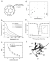
Figure 1
Figure 1
Evidence of fullerenol binding to Ub and Ub2. a) Chemical structure of the fullerenol particle used in this study. b) Fluorescence emission spectra of Ub (Tyr59, excitation at 280 nm) in the presence of fullerenol. c) Normalized maximum emission fluorescence measured on Ub at increasing fullerenol concentration (lines serve to guide the eye). d) Rotational correlation time for Ub and Ub2 in the presence of fullerenol, determined from 15N spin relaxation measurements. e) Saturation profile of the 1D HN envelope of Ub by transfer of 15N saturation from the NP-bound state. The inset highlights the intensity differences measured for Ub alone and in the presence of NPs. Errors in intensities were estimated from spectral noise and are about the size of the symbols. f) Cartoon representation of Ub with secondary structure elements indicated.
Figure 2
Figure 2
NMR signal perturbations of Ub in the presence of fullerenol. a) Overlaid regions of 1H,15N-HSQC NMR spectra collected on 15N-Ub in the absence (dark grey) and presence (red) of fullerenol at [fullerenol]/[Ub] = 1.5. b) CSPs measured at the [fullerenol]/[Ub] molar ratios reported in the legend. Secondary structure elements are reported on top of the panel. c) CSPs and intensity perturbations measured at [fullerenol]/[Ub] = 1.5. Intensity perturbation was computed as: (I0-Ii)/I0, where Ii is the signal intensity at titration step i, and I0 is the signal intensity of free Ub. d) Representative CSP-based binding isotherms.
Figure 3
Figure 3
NMR signal perturbations of Ub2 in the presence of fullerenol. a, b) Overlaid regions of 1H,15N-HSQC NMR spectra collected in the absence (dark grey) and presence (red) of fullerenol at [fullerenol]/[Ub2] = 1.5. In the schematic representation of Ub2 on the right of each spectrum, the 15N-labelled Ub unit is coloured in grey, the unlabelled unit is displayed in white. c, d) CSPs measured at the [fullerenol]/[Ub2] values indicated in the legend. e, f) CSPs and intensity perturbations measured at [fullerenol]/[Ub2] = 0.75. g,h) CSPs and intensity perturbations measured at [fullerenol]/[Ub2] = 1.5. Panels a, c, e, g display data measured on Ub2(15N-D) while the data in panels b, d, f, h correspond to Ub2(15N-P).
Figure 4
Figure 4
Mapping of the fullerenol binding surface on ubiquitin. a) Mapping of Ub residues (yellow sticks) displaying greater-than-average intensity perturbations after addition of fullerenol ([fullerenol]/[Ub] = 1.5); key interface residues are indicated; asterisks indicate the sites of covalent modification involved in formation of Lys48-linked chains; the shown structure is 1UBQ. b) Ub molecular surface with the fullerenol-binding epitope painted yellow. c) Mapping of Ub2 residues (yellow sticks) displaying greater-than-average intensity perturbations after addition of fullerenol ([fullerenol]/[Ub2] = 1.5); yellow colored residues in the proximal Ub correspond to an intensity attenuation > 0.95, the residues that exhibited an intensity attenuation between 0.93 and 0.95 are shown in gray sticks; the distal (carrying free Lys48) and proximal (carrying free C-terminus) units are schematically depicted in the bottom; the figure corresponds to the ‘open’ solution structure 2PE9. d) Molecular surface of Ub2 with the fullerenol-binding epitope painted yellow. A fullerenol molecule is shown between panels b and d in scale with all proteins displayed in the figure.
Figure 5
Figure 5
PolyUb chain formation monitored by SDS-PAGE. Ub and its mutants Ub(K48R) and Ub(D77) have a molecular weight of ~8.5 kDa. Ub2, Ub3, and Ub4 have molecular weights of 17, 25.5, and 34 kDa, respectively. GST-E2 has a molecular weight of about 50 kDa. Addition of fullerenol at the same molar concentration as Ub abolished polyUb chain formation. Results for Ub(K48R) and Ub(D77) are shown as these mutants were used for Ub2 production in all experiments described in this work. These mutations prevent formation of chains longer than the dimer.

References

    1. Linse S, Cabaleiro-Lago C, Xue W-F, Lynch I, Lindman S, Thulin E, Radford SE, Dawson KA. Proc. Natl. Acad. Sci. U. S. A. 2007;104:8691–8696. - PMC - PubMed
    1. Nel A, Xia T, Mädler L, Li N. Science. 2006;311:622–627. - PubMed
    1. Saptarshi SR, Duschl A, Lopata AL. J. Nanobiotechnology. 2013;11:26. - PMC - PubMed
    1. Aguilar RC, Wendland B. Curr. Opin. Cell Biol. 2003;15:184–190. - PubMed
    1. Pickart CM, Fushman D. Curr. Opin. Chem. Biol. 2004;8:610–616. - PubMed

Publication types

MeSH terms

LinkOut - more resources